A Successful Control of the Intraoperative Bleeding from McConnell’s Artery during Fully Endoscopic Resection of Planum Sphenoidale Meningioma Using Bone Chip and Bioglue : A Case Report
- Affiliations
-
- 1Department of Neurosurgery, General Hospital Bamberg, Bamberg, Germany
- 2Department of Neurosurgery, University Hospital Tuebingen, Tuebingen, Germany
- 3Department of Neurological Surgery, Houston Methodist Hospital, Houston, TX, USA
- 4Department of Neurosurgery, University Medical Centre Ljubljana, Ljubljana, Slovenia
- KMID: 2565279
- DOI: http://doi.org/10.3340/jkns.2024.0143
Abstract
- The endoscopic transsphenoidal approach is a common approach used in skull base neurosurgery to reach the sellar region. One of the intraoperative risks of this approach is intraoperative bleeding out of the carotid artery. Gentle drilling can prevent carotid artery injury. However, injury to smaller branches, such as the McConnell’s capsular artery, which is located within the surgical corridor, is more difficult to prevent. If such an injury is within the junction to the main trunk of the carotid artery, there will be a small circular defect in this area. This can result in massive blood loss and should be closed surgically immediately. We describe a clinical case of intraoperative bleeding from the McConnell’s artery originating from the carotid arterial segment (C4) in a 78-year-old female patient operated on for planum sphenoidale meningioma via endoscopic transsphenoidal approach, as well as provide a technical note on a possible technique for bleeding control in such cases. Pinpoint carotid bleeding as a result of intraoperative injury can be stopped by wedging a bone fragment in the carotid canal and fixing it in that position with histoacryl glue at the defect site.
Figure
-
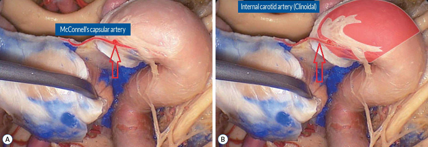
Fig. 1. McConnell’s artery is marked with red arrow (A and B). Reused from Rhoton [13].
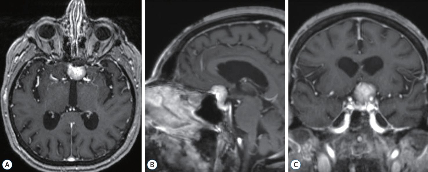
Fig. 2. The preoperative contrast-enhanced T1-weighted MRI scans in the axial (A), sagittal (B), and coronal (C) plans show planum sphenoidale meningioma with left optic nerve compression.
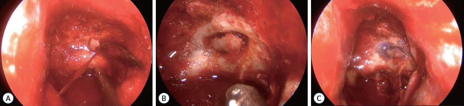
Fig. 3. The intraoperative photos show bleeding from the McConnell’s capsular artery (A) and a fixed bone fragment at the defect site (B and C).
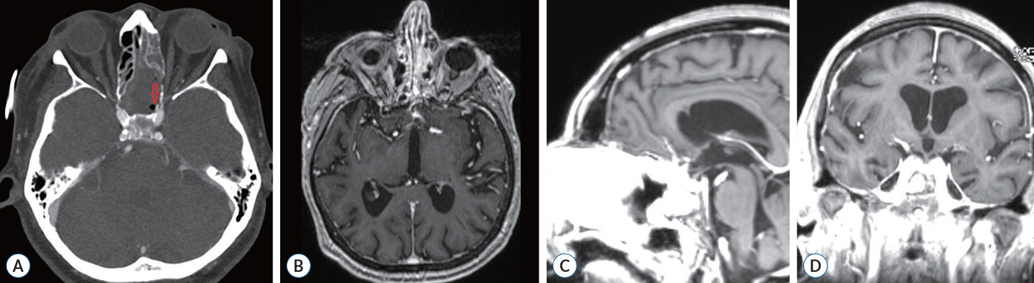
Fig. 4. The postoperative computed tomography (CT) and magnetic resonance imaging (MRI) scans. A : The CT angiography scans immediately after the surgery did not show pseudoaneurysms or active carotid bleeding (inserted bone chip is marked with red arrow). The postoperative contrast-enhanced T1-weighted MRI scans in the axial (B), sagittal (C), and coronal (D) plans made 3 months after the surgery show no tumor recurrence.
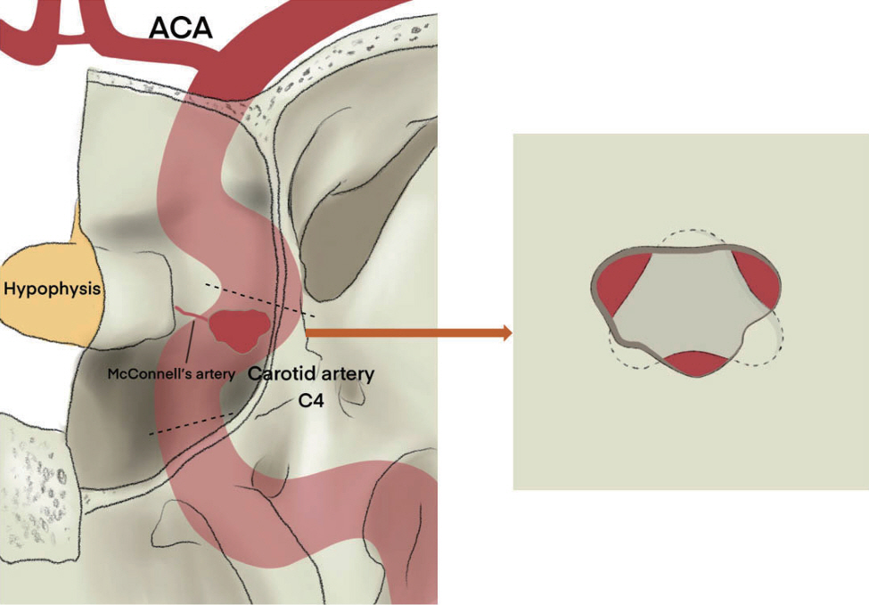
Fig. 5. Schematic illustration of closing the defect in the carotid arterial wall by wedging a bone fragment in the carotid canal between the carotid artery and the surrounding bone. Courtesy of Palina Andreyeva. ACA : anterior cerebral artery.
Reference
-
References
1. Cavallo LM, Briganti F, Cappabianca P, Maiuri F, Valente V, Tortora F, et al. Hemorrhagic vascular complications of endoscopic transsphenoidal surgery. Minim Invasive Neurosurg. 47:145–150. 2004.
Article2. Charalampaki P, Ayyad A, Kockro RA, Perneczky A. Surgical complications after endoscopic transsphenoidal pituitary surgery. J Clin Neurosci. 16:786–789. 2009.
Article3. Ciric I, Ragin A, Baumgartner C, Pierce D. Complications of transsphenoidal surgery: results of a national survey, review of the literature, and personal experience. Neurosurgery. 40:225–236. discussion 236-237. 1997.
Article4. Debus ES, Grundmann RT. Acute carotid part 3: artery injury. Gefasschirurgie. 25:356–363. 2020.5. Ferrareze Nunes C, Beer-Furlan A, Doglietto F, Carrau RL, Prevedello DM. The McConnell’s capsular arteries and their relevance in endoscopic endonasal approach to the sellar region. Oper Neurosurg (Hagerstown). 14:171–177. 2018.
Article6. Gardner PA, Snyderman CH, Fernandez-Miranda JC, Jankowitz BT. Management of major vascular injury during endoscopic endonasal skull base surgery. Otolaryngol Clin North Am. 49:819–828. 2016.
Article7. Gardner PA, Tormenti MJ, Pant H, Fernandez-Miranda JC, Snyderman CH, Horowitz MB. Carotid artery injury during endoscopic endonasal skull base surgery: incidence and outcomes. Neurosurgery. 73(2 Suppl Operative):ons261–ons269. discussion ons269-ons270. 2013.8. Kalinin PL, Sharipov OI, Shkarubo AN, Fomichev DV, Kutin MA, Alekseev SN, et al. Damage to the cavernous segment of internal carotid artery in transsphenoidal endoscopic removal of pituitary adenomas (report of 4 cases). Zh Vopr Neirokhir Im N N Burdenko. 77:28–37. 2013.9. Li C, Zhu H, Zong X, Wang X, Gui S, Zhao P, et al. Experience of trans-nasal endoscopic surgery for pituitary tumors in a single center in China : surgical results in a cohort of 2032 patients, operated between 2006 and 2018. Clin Neurol Neurosurg. 197:106176. 2020.10. Oskouian RJ, Kelly DF, Laws ERJ Jr. Vascular injury and transsphenoidal surgery. Front Horm Res. 34:256–278. 2006.
Article11. Padhye V, Valentine R, Wormald PJ. Management of carotid artery injury in endonasal surgery. Int Arch Otorhinolaryngol. 18(Suppl 2):S173–S178. 2014.
Article12. Raymond J, Hardy J, Czepko R, Roy D. Arterial injuries in transsphenoidal surgery for pituitary adenoma; the role of angiography and endovascular treatment. AJNR Am J Neuroradiol. 18:655–665. 1997.13. Rhoton AL Jr. Cranial Anatomy and Surgical Approach. Philadelphia: Lippincott Williams & Wilkins;2003.14. Romero ADCB, Lal Gangadharan J, Bander ED, Gobin YP, Anand VK, Schwartz TH. Managing arterial injury in endoscopic skull base surgery: case series and review of the literature. Oper Neurosurg (Hagerstown). 13:138–149. 2017.
Article15. Rowan NR, Turner MT, Valappil B, Fernandez-Miranda JC, Wang EW, Gardner PA, et al. Injury of the carotid artery during endoscopic endonasal surgery: surveys of skull base surgeons. J Neurol Surg B Skull Base. 79:302–308. 2018.
Article16. Sharma RK, Irace AL, Overdevest JB, Gudis DA. Carotid artery injury in endoscopic endonasal surgery: risk factors, prevention, and management. World J Otorhinolaryngol Head Neck Surg. 8:54–60. 2022.
Article17. Valentine R, Padhye V, Wormald PJ. Management of arterial injury during endoscopic sinus and skull base surgery. Curr Opin Otolaryngol Head Neck Surg. 24:170–174. 2016.
Article18. Wang WH, Lieber S, Lan MY, Wang EW, Fernandez-Miranda JC, Snyderman CH, et al. Nasopharyngeal muscle patch for the management of internal carotid artery injury in endoscopic endonasal surgery. J Neurosurg. 133:1382–1387. 2020.
Article19. Zhang Y, Tian Z, Li C, Liu J, Zhang Y, Yang X, et al. A modified endovascular treatment protocol for iatrogenic internal carotid artery injuries following endoscopic endonasal surgery. J Neurosurg. 132:343–350. 2020.
Article
- Full Text Links
-
- Actions
-
Cited
- CITED
-
- Close
- Share
-
- Similar articles
-
- Convexity Meningioma En Plaque Presenting with Diffuse Hyperostosis of the Skull
- Experience with 5-Aminolevulinic Acid in Fluorescence-Guided Resection of a Deep Sylvian Meningioma
- Modified Anterior Craniofacial Osteotomy Using Partial Nasal Bone Division and Reconstruction in Frontoethmoidal Sinus Meningioma
- A Case of Sporadic Suprasellar Hemangioblastoma Mimicking Meningioma
- Visual Recovery with Surgical Managment of Suprasellar Meningioma
