Korean J Sports Med.
2024 Dec;42(4):289-295. 10.5763/kjsm.2024.42.4.289.
Lower Extremity Biomechanical Comparison Analysis of Single Leg Drop Landing among Normal Foot and Flat Foot
- Affiliations
-
- 1Department of Physical Education, Graduate School of Pukyong National University, Busan, Korea
- 2Major of Marine Sports, Division of Smart Healthcare, Pukyong National University, Busan, Korea
- KMID: 2561757
- DOI: http://doi.org/10.5763/kjsm.2024.42.4.289
Abstract
- Purpose
The purpose of this study is to compare and analyze the differences in biomechanical function and energy absorption of the lower extremity in the sagittal plane when single leg drop landing between groups with flat foot and normal foot.
Methods
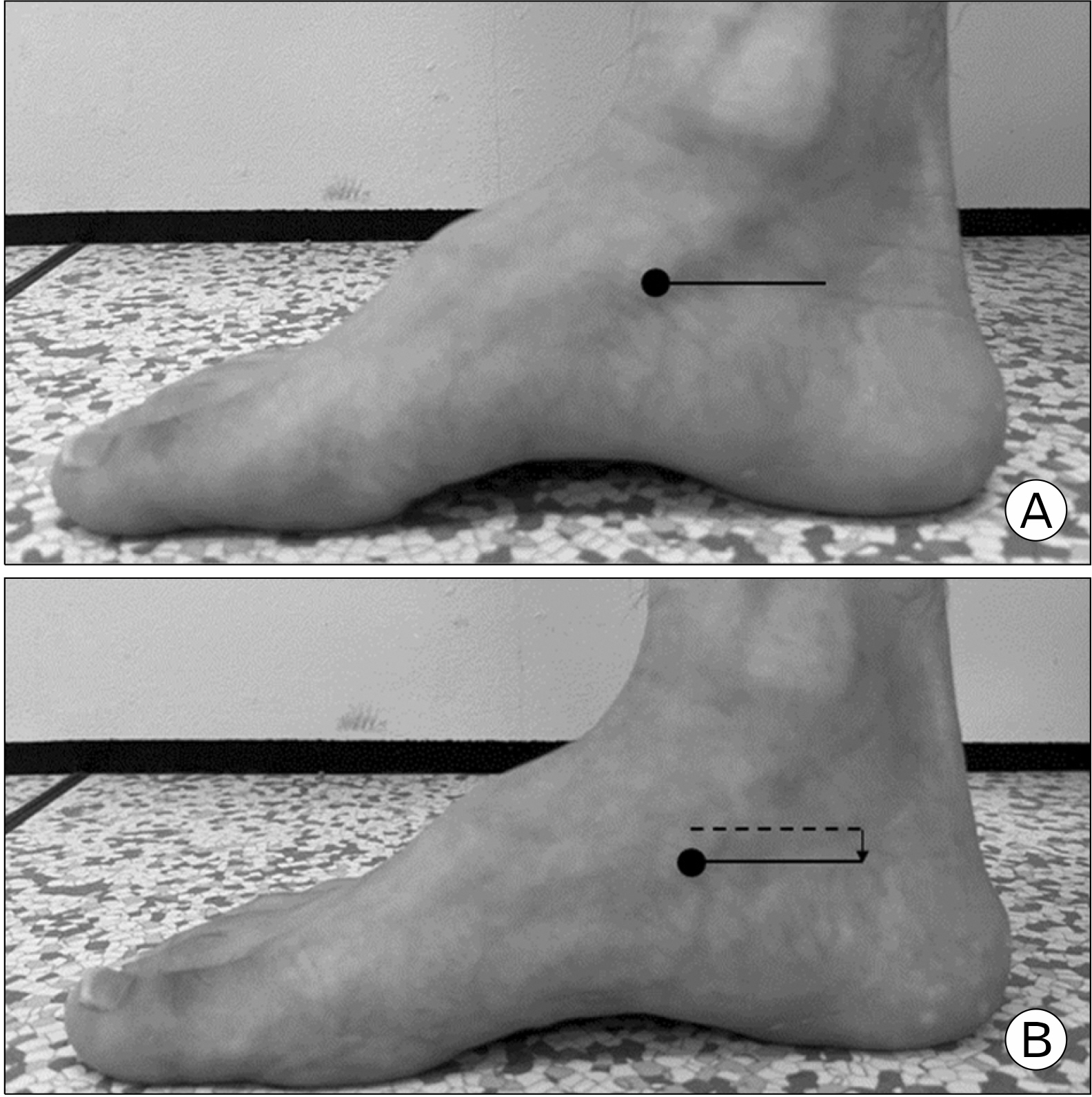
Twenty-eight healthy men in their 20s were classified into 13 with flat foot and 15 with normal foot through evaluation of navicular drop test. Using a motion analysis system, loading rate (N/sec), peak vertical ground reaction force (N/body weight [BW]), sagittal plane hip, knee, and ankle joint range of motion (°), peak moment (Nm/BW), peak joint power (W/BW) and peak joint work (J/BW) were calculated and analyzed during single leg drop landing.
Results
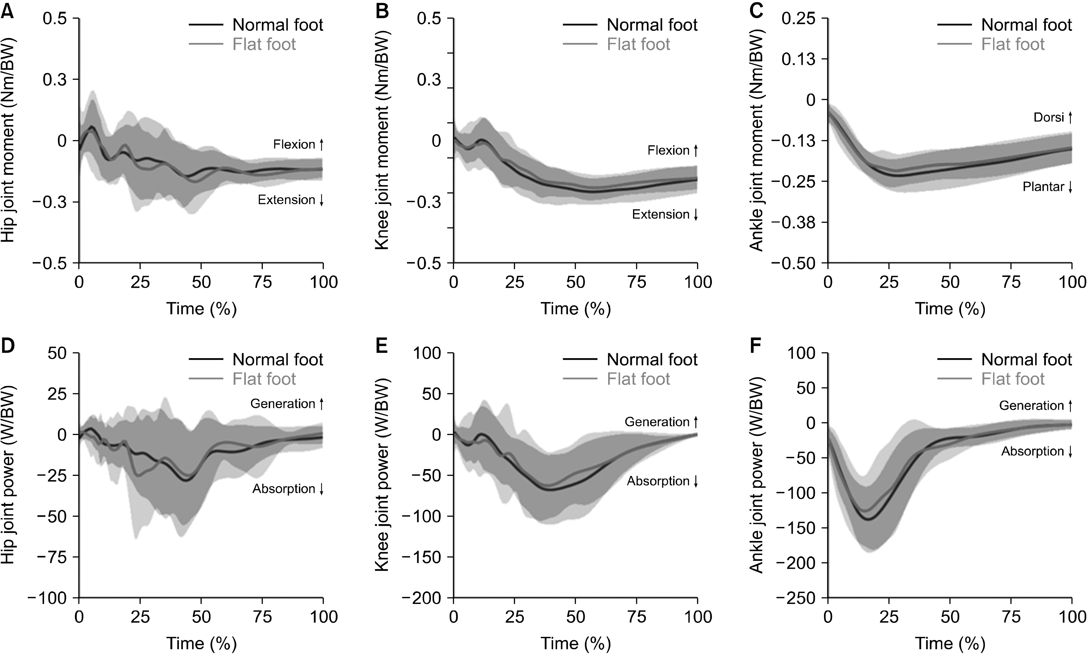
During single leg drop landing, the flat foot and normal foot groups showed no significant differences in loading rate, peak vertical ground reaction force, hip and knee joint range of motion, peak knee and ankle joint moment, peak joint power, and peak joint work (p> 0.05). However, the flat foot group showed greater ankle range of motion and peak hip joint flexion moment compared to the normal foot group (p=0.040 and p=0.018, respectively).
Conclusion
The flat foot group shows sagittal plane landing mechanics that are different from the normal foot group during single leg drop landing and appears to try to distribute shock by relying on the distal joint compared to the normal foot group.
Figure
Reference
-
1. Root ML, Orien WP, Weed JH. Normal and abnormal function of the foot. Clinical Biomechanics Corp.;1977.2. Fiolkowski P, Brunt D, Bishop M, Woo R, Horodyski M. 2003; Intrinsic pedal musculature support of the medial longitudinal arch: an electromyography study. J Foot Ankle Surg. 42:327–33. DOI: 10.1053/j.jfas.2003.10.003. PMID: 14688773.3. Tang CY, Ng KH, Lai J. 2020; Adult flatfoot. BMJ. 368:m295. DOI: 10.1136/bmj.m295. PMID: 32094144.4. Arangio GA, Phillippy DC, Xiao D, Gu WK, Salathe EP. 2000; Subtalar pronation: relationship to the medial longitudinal arch loading in the normal foot. Foot Ankle Int. 21:216–20. DOI: 10.1177/107110070002100306. PMID: 10739152.5. Levinger P, Gilleard W. 2007; Tibia and rearfoot motion and ground reaction forces in subjects with patellofemoral pain syndrome during walking. Gait Posture. 25:2–8. DOI: 10.1016/j.gaitpost.2005.12.015. PMID: 16483778.6. Kothari A, Dixon PC, Stebbins J, Zavatsky AB, Theologis T. 2016; Are flexible flat feet associated with proximal joint problems in children? Gait Posture. 45:204–10. DOI: 10.1016/j.gaitpost.2016.02.008. PMID: 26979907.7. Maharaj JN, Cresswell AG, Lichtwark GA. 2017; Foot structure is significantly associated to subtalar joint kinetics and mechanical energetics. Gait Posture. 58:159–65. DOI: 10.1016/j.gaitpost.2017.07.108. PMID: 28783556.8. Ng JW, Chong LJ, Pan JW, Lam WK, Ho M, Kong PW. 2021; Effects of foot orthosis on ground reaction forces and perception during short sprints in flat-footed athletes. Res Sports Med. 29:43–55. DOI: 10.1080/15438627.2020.1755673. PMID: 32326755.9. Tong JW, Kong PW. 2013; Association between foot type and lower extremity injuries: systematic literature review with meta-analysis. J Orthop Sports Phys Ther. 43:700–14. DOI: 10.2519/jospt.2013.4225. PMID: 23756327.10. McNitt-Gray JL. 1991; Kinematics and impulse characteristics of drop landings from three heights. J Appl Biomech. 7:201–24. DOI: 10.1123/ijsb.7.2.201.11. Stuelcken MC, Mellifont DB, Gorman AD, Sayers MG. 2016; Mechanisms of anterior cruciate ligament injuries in elite women's netball: a systematic video analysis. J Sports Sci. 34:1516–22. DOI: 10.1080/02640414.2015.1121285. PMID: 26644060.12. Sung PS, Zipple JT, Andraka JM, Danial P. 2017; The kinetic and kinematic stability measures in healthy adult subjects with and without flat foot. Foot (Edinb). 30:21–6. DOI: 10.1016/j.foot.2017.01.010. PMID: 28257946.13. Fu FQ, Wang S, Shu Y, Li JS, Popik S, Gu YD. 2016; A comparative biomechanical analysis the vertical jump between flatfoot and normal foot. J Biomim Biomater Biomed Eng. 28:26–35. DOI: 10.4028/www.scientific.net/JBBBE.28.26.14. Choi JH, An HJ, Yoo KT. 2012; Comparison of the loading rate and lower limb angles on drop-landing between a normal foot and flatfoot. J Phys Ther Sci. 24:1153–7. DOI: 10.1589/jpts.24.1153.15. Devita P, Skelly WA. 1992; Effect of landing stiffness on joint kinetics and energetics in the lower extremity. Med Sci Sports Exerc. 24:108–15. DOI: 10.1249/00005768-199201000-00018. PMID: 1548984.16. Cote KP, Brunet ME, Gansneder BM, Shultz SJ. 2005; Effects of pronated and supinated foot postures on static and dynamic postural stability. J Athl Train. 40:41–6.17. Maniar N, Schache AG, Pizzolato C, Opar DA. 2020; Muscle contributions to tibiofemoral shear forces and valgus and rotational joint moments during single leg drop landing. Scand J Med Sci Sports. 30:1664–74. DOI: 10.1111/sms.13711. PMID: 32416625.18. McNitt-Gray JL. 1993; Kinetics of the lower extremities during drop landings from three heights. J Biomech. 26:1037–46. DOI: 10.1016/S0021-9290(05)80003-X. PMID: 8408086.19. Franco AH. 1987; Pes cavus and pes planus: analyses and treatment. Phys Ther. 67:688–94. DOI: 10.1093/ptj/67.5.688. PMID: 3575426.20. Chang JS, Kwon YH, Choi JH, Lee HS. 2012; Gender differences in lower extremity kinematics and kinetics of the vertical ground reaction force peak in drop-landing by flatfooted subjects. J Phys Ther Sci. 24:267–70. DOI: 10.1589/jpts.24.267.21. Adouni M, Shirazi-Adl A, Marouane H. 2016; Role of gastrocnemius activation in knee joint biomechanics: gastrocnemius acts as an ACL antagonist. Comput Methods Biomech Biomed Engin. 19:376–85. DOI: 10.1080/10255842.2015.1032943. PMID: 25892616.22. Hewett TE, Myer GD, Ford KR, et al. 2005; Biomechanical measures of neuromuscular control and valgus loading of the knee predict anterior cruciate ligament injury risk in female athletes: a prospective study. Am J Sports Med. 33:492–501. DOI: 10.1177/0363546504269591. PMID: 15722287.23. Arampatzis A, Morey-Klapsing G, Brüggemann GP. 2003; The effect of falling height on muscle activity and foot motion during landings. J Electromyogr Kinesiol. 13:533–44. DOI: 10.1016/S1050-6411(03)00059-2. PMID: 14573368.24. Hargrave MD, Carcia CR, Gansneder BM, Shultz SJ. 2003; Subtalar pronation does not influence impact forces or rate of loading during a single-leg landing. J Athl Train. 38:18–23.25. Behling AV, Nigg BM. 2020; Relationships between the foot posture index and static as well as dynamic rear foot and arch variables. J Biomech. 98:109448. DOI: 10.1016/j.jbiomech.2019.109448. PMID: 31677779.
- Full Text Links
- Actions
-
Cited
- CITED
-
- Close
- Share
- Similar articles
-
- Comparisons of Vastus Medialis and Vastus Lateralis Muscle Activities according to Different Heights during Drop Landing in Flatfooted Adults
- Surgical Correction of Foot Drop in Leprosy
- Biomechanic Analysis of Lower Extremities in Children and Teenagers with Pes Planus
- Acute Compartment Syndrome of the Lower Leg and Foot
- Foot Drop after Anterior Cervical Discectomy and Fusion: A Case Report


