J Yeungnam Med Sci.
2024 Oct;41(4):296-299. 10.12701/jyms.2024.00577.
Myxoid lipoma in the perioral mandibular region: two case reports
- Affiliations
-
- 1Department of Dentistry, Yeungnam University College of Medicine, Daegu, Korea
- 2Department of Oral and Maxillofacial Surgery, School of Dentistry, Pusan National University, Yangsan, Korea
- 3Dental and Life Science Institute, Pusan National University, Busan, Korea
- KMID: 2560490
- DOI: http://doi.org/10.12701/jyms.2024.00577
Abstract
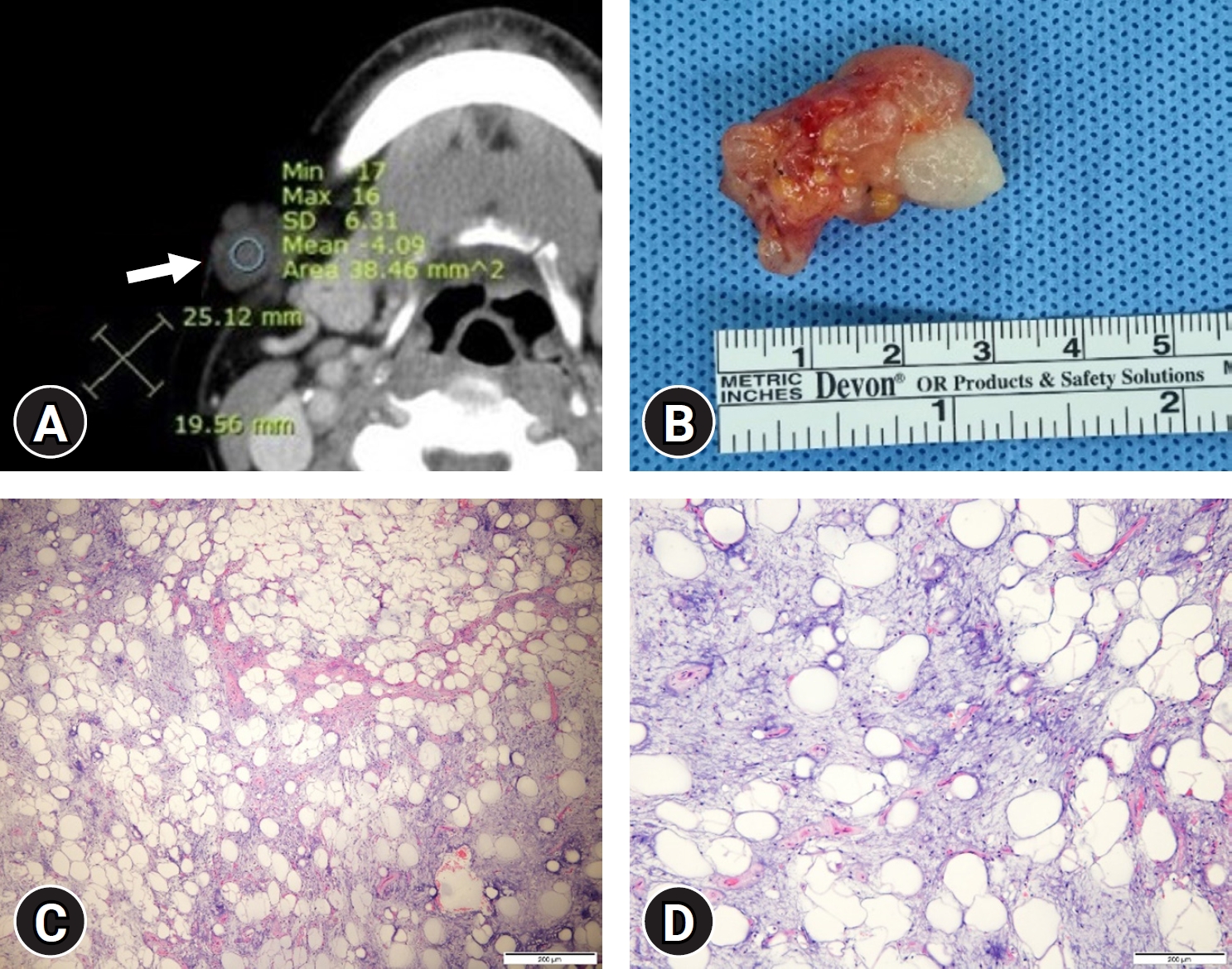
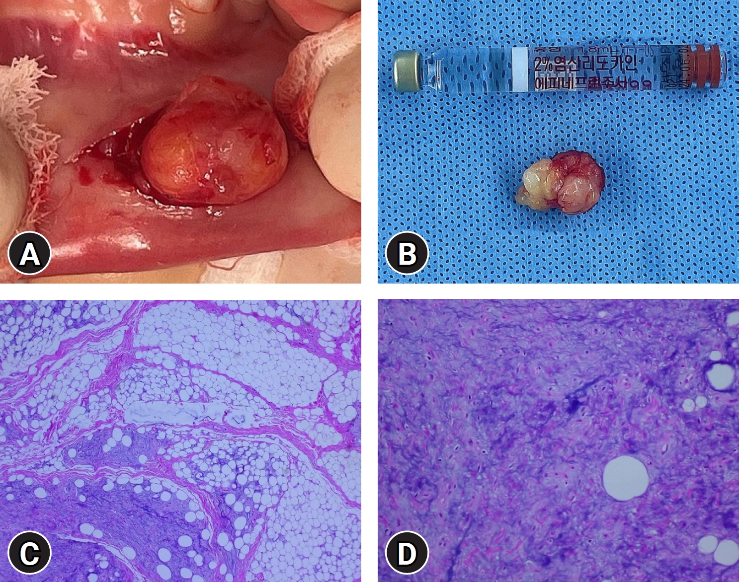
- Lipomas are one of the most common mesenchymal tumors in the human body, exhibiting a heightened prevalence between the ages of 40 and 60 years. However, primary intraoral lipomas are rare. Myxoid lipoma, which is characterized by abundant mucoid components, is a particularly rare histological subtype of lipoma. This study presents two cases of myxoid lipoma that occurred outside the common age range for occurrence, one in the right submandibular area of a 67-year-old male and the other in the lower lip of a 3-year-old child. Through these case reports, the aim was to introduce myxoid lipoma, a rare subtype affecting facial areas, and provide a brief review to assist in the differential diagnosis, emphasizing the importance of pathological assessment. Even in age groups and anatomical locations not typically associated with lipomas, it is crucial to emphasize the necessity of careful evaluation.
Keyword
Figure
Reference
-
References
1. Kolb L, Yarrarapu SN, Ameer MA, Rosario-Collazo JA. Lipoma [updated 2023 Aug 8]. In: StatPearls [Internet]. Treasure Island (FL): StatPearls Publishing;2024. [cited 2024 Jun 12]. https://www.ncbi.nlm.nih.gov/books/NBK507906/.2. Ono S, Rana M, Takechi M, Ogawa I, Okui G, Mitani Y, et al. Myxolipoma in the tongue - a clinical case report and review of the literature. Head Neck Oncol. 2011; 3:50.3. Curien R, Aldosa J, Beurey P, Bravetti P. Posttraumatic myxoid lipoma of the lower lip mimicking a mucocele. Med Buccale Chir Buccale. 2012; 18:233–4.4. Chen SY, Fantasia JE, Miller AS. Myxoid lipoma of oral soft tissue. A clinical and ultrastructural study. Oral Surg Oral Med Oral Pathol. 1984; 57:300–7.5. Studart-Soares EC, Costa FW, Sousa FB, Alves AP, Osterne RL. Oral lipomas in a Brazilian population: a 10-year study and analysis of 450 cases reported in the literature. Med Oral Patol Oral Cir Bucal. 2010; 15:e691–6.6. Ausband JR, Harrill JA, Pautler EE. Myxolipoma of the base of the tongue; report of a case. Ann Otol Rhinol Laryngol. 1953; 62:1039–43.7. Allon I, Kaplan I, Gal G, Chaushu G, Allon DM. The clinical characteristics of benign oral mucosal tumors. Med Oral Patol Oral Cir Bucal. 2014; 19:e438–43.8. Copcu E, Sivrioglu NS. Posttraumatic lipoma: analysis of 10 cases and explanation of possible mechanisms. Dermatol Surg. 2003; 29:215–20.


