Korean J Orthod.
2024 Sep;54(5):316-324. 10.4041/kjod23.261.
Dentoalveolar effects of open-bite correction with the dual action vertical intra-arch technique: A finite element analysis
- Affiliations
-
- 1Division of Orthodontics, Federal University of Rio Grande do Sul, Porto Alegre, Brazil
- 2Division of Technologies for Production and Health, Renato Archer Information Technology Center, Campinas, Brazil
- KMID: 2559820
- DOI: http://doi.org/10.4041/kjod23.261
Abstract
Objective
To evaluate tooth displacement and periodontal stress generated by the dual action vertical intra-arch technique (DAVIT) for open-bite correction using three-dimensional finite element analysis.
Methods
A three-dimensional model of the maxilla was created by modeling the cortical bone, cancellous bone, periodontal ligament, and teeth from the second molar to the central incisor of a hemiarch. All orthodontic devices were designed using specific software to reproduce their morpho-dimensional characteristics, and their physical properties were determined using Young’s modulus and Poisson’s coefficient of each material. A linear static simulation was performed to analyze the tooth displacements (mm) and maximum stresses (Mpa) induced in the periodontal ligament by the posterior intrusion and anterior extrusion forces generated by the DAVIT.
Results
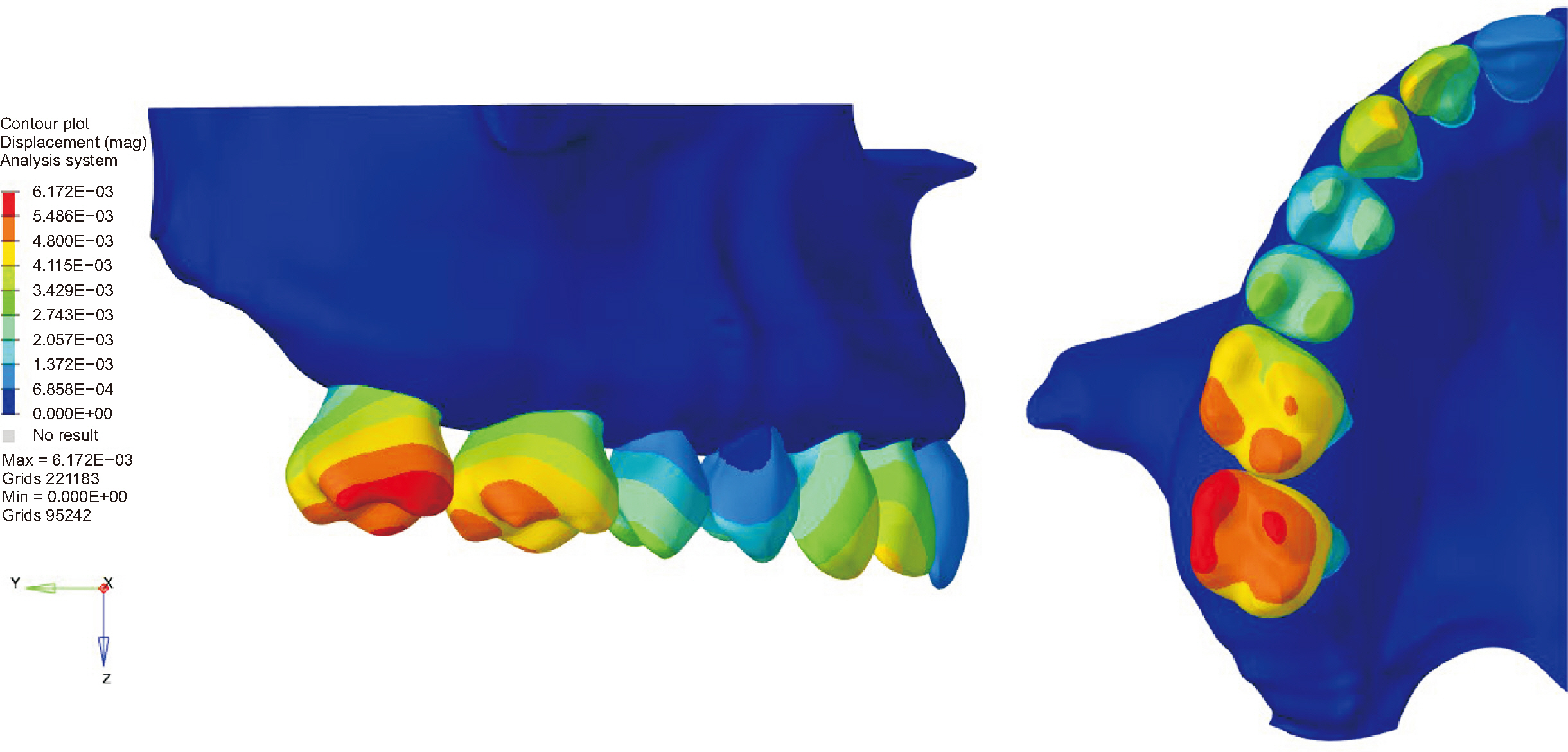
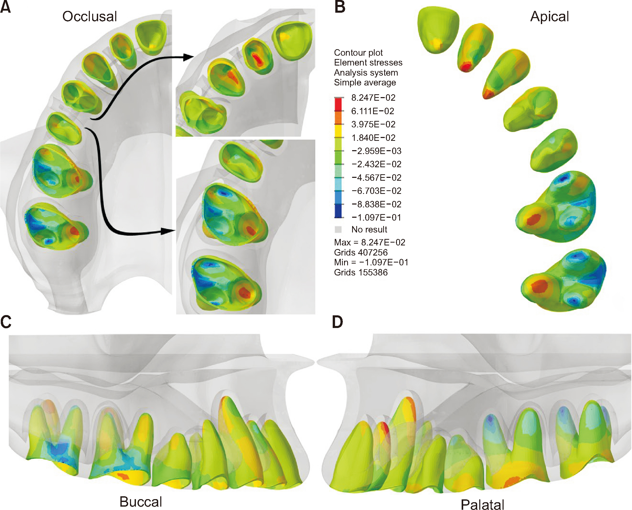
The first and second molars showed the greatest intrusion, whereas the canines and lateral incisors showed the greatest extrusion displacement. A neutral zone of displacement corresponding to the fulcrum of occlusal plane rotation was observed in the premolar region. Buccal tipping of the molars and lingual tipping of the anterior teeth occurred with intrusion and extrusion, respectively. Posterior intrusion generated compressive stress at the apex of the buccal roots and furcation of the molars, while anterior extrusion generated tensile stress at the apex and apical third of the palatal root surface of the incisors and canines.
Conclusions
DAVIT mechanics produced a set of beneficial effects for open-bite correction, including molar intrusion, extrusion and palatal tipping of the anterior teeth, and occlusal plane rotation with posterior teeth uprighting.
Figure
Reference
-
1. Alsafadi AS, Alabdullah MM, Saltaji H, Abdo A, Youssef M. 2016; Effect of molar intrusion with temporary anchorage devices in patients with anterior open bite: a systematic review. Prog Orthod. 17:9. https://doi.org/10.1186/s40510-016-0122-4. DOI: 10.1186/s40510-016-0122-4. PMID: 26980200. PMCID: PMC4803715. PMID: 2bfea0e6693d479088c4e96e5a695b05.2. Beane RA Jr. 1999; Nonsurgical management of the anterior open bite: a review of the options. Semin Orthod. 5:275–83. https://doi.org/10.1016/s1073-8746(99)80021-8. DOI: 10.1016/S1073-8746(99)80021-8.3. Hart TR, Cousley RR, Fishman LS, Tallents RH. 2015; Dentoskeletal changes following mini-implant molar intrusion in anterior open bite patients. Angle Orthod. 85:941–8. https://doi.org/10.2319/090514-625.1. DOI: 10.2319/090514-625.1. PMID: 25531420. PMCID: PMC8612037.4. Marzouk ES, Kassem HE. 2016; Evaluation of long-term stability of skeletal anterior open bite correction in adults treated with maxillary posterior segment intrusion using zygomatic miniplates. Am J Orthod Dentofacial Orthop. 150:78–88. https://doi.org/10.1016/j.ajodo.2015.12.014. DOI: 10.1016/j.ajodo.2015.12.014.5. Park YC, Lee HA, Choi NC, Kim DH. 2008; Open bite correction by intrusion of posterior teeth with miniscrews. Angle Orthod. 78:699–710. https://doi.org/10.2319/0003-3219(2008)078[0699:OBCBIO]2.0.CO;2. DOI: 10.2319/0003-3219(2008)078[0699:OBCBIO]2.0.CO;2.6. Scheffler NR, Proffit WR, Phillips C. 2014; Outcomes and stability in patients with anterior open bite and long anterior face height treated with temporary anchorage devices and a maxillary intrusion splint. Am J Orthod Dentofacial Orthop. 146:594–602. https://doi.org/10.1016/j.ajodo.2014.07.020. DOI: 10.1016/j.ajodo.2014.07.020.7. Atsawasuwan P, Hohlt W, Evans CA. 2015; Nonsurgical approach to class I open-bite malocclusion with extrusion mechanics: a 3-year retention case report. Am J Orthod Dentofacial Orthop. 147:499–508. https://doi.org/10.1016/j.ajodo.2014.04.024. DOI: 10.1016/j.ajodo.2014.04.024.8. Isaacson RJ, Lindauer SJ. 2001; Closing anterior open bites: the extrusion arch. Semin Orthod. 7:34–41. https://doi.org/10.1053/sodo.2001.21064. DOI: 10.1053/sodo.2001.21064.9. Janson G, Valarelli FP, Henriques JF, de Freitas MR, Cançado RH. 2003; Stability of anterior open bite nonextraction treatment in the permanent dentition. Am J Orthod Dentofacial Orthop. 124:265–76. quiz 340https://doi.org/10.1016/s0889-5406(03)00449-9. DOI: 10.1016/S0889-5406(03)00449-9.10. Lindauer SJ, Isaacson RJ. 1995; One-couple orthodontic appliance systems. Semin Orthod. 1:12–24. https://doi.org/10.1016/s1073-8746(95)80084-0. DOI: 10.1016/S1073-8746(95)80084-0.11. Cruz-Escalante MA, Aliaga-Del Castillo A, Soldevilla L, Janson G, Yatabe M, Zuazola RV. 2017; Extreme skeletal open bite correction with vertical elastics. Angle Orthod. 87:911–23. https://doi.org/0.2319/042817-287.1. DOI: 10.2319/042817-287.1. PMID: 28895751. PMCID: PMC8317572.12. Gudhimella S, Gandhi V, Schiro NL, Janakiraman N. 2022; Management of anterior open bite and skeletal class II hyperdivergent patient with clear aligner therapy. Turk J Orthod. 35:139–49. https://doi.org/10.5152/TurkJOrthod.2022.21053. DOI: 10.5152/TurkJOrthod.2022.21053. PMID: 35788439. PMCID: PMC9317343. PMID: 2b73f7eef3d4443a85f2374075d116f5.13. Kaku M, Kawai A, Koseki H, Abedini S, Kawazoe A, Sasamoto T, et al. 2009; Correction of severe open bite using miniscrew anchorage. Aust Dent J. 54:374–80. https://doi.org/10.1111/j.1834-7819.2009.01166.x. DOI: 10.1111/j.1834-7819.2009.01166.x. PMID: 20415938.14. Kim YH. 1987; Anterior openbite and its treatment with multiloop edgewise archwire. Angle Orthod. 57:290–321. https://doi.org/10.1043/0003-3219(1987)057<0290:AOAITW>2.0.CO;2.15. Kim YH, Han UK, Lim DD, Serraon ML. 2000; Stability of anterior openbite correction with multiloop edgewise archwire therapy: a cephalometric follow-up study. Am J Orthod Dentofacial Orthop. 118:43–54. https://doi.org/10.1067/mod.2000.104830. DOI: 10.1067/mod.2000.104830.16. Barros SE, Chiqueto K, Janson G, Janson M. 2022; Dual action vertical intra-arch technique. J Clin Orthod. 56:666–76. https://pubmed.ncbi.nlm.nih.gov/37158769/. DOI: 10.4041/kjod23.261.17. Barros SE, Chiqueto K, Heck B, Faria J, Ferreira E, Janson M. 2023; Dual-action vertical intraarch technique: a multifocal technique for open bite correction. AJO-DO Clin Companion. 3:286–95. https://doi.org/10.1016/j.xaor.2023.06.001. DOI: 10.1016/j.xaor.2023.06.001.18. Barros SE, Faria J, Jaramillo Cevallos K, Chiqueto K, Machado L, Noritomi P. 2022; Torqued and conventional cantilever for uprighting mesially impacted molars: a 3-dimensional finite element analysis. Am J Orthod Dentofacial Orthop. 162:e203–15. https://doi.org/10.1016/j.ajodo.2022.07.014. DOI: 10.1016/j.ajodo.2022.07.014. PMID: 35999156.19. Heboyan A, Lo Giudice R, Kalman L, Zafar MS, Tribst JPM. 2022; Stress distribution pattern in zygomatic implants supporting different superstructure materials. Materials (Basel). 15:4953. https://doi.org/10.3390/ma15144953. DOI: 10.3390/ma15144953. PMID: 35888420. PMCID: PMC9323759. PMID: f4781cd82f5d4bc78eb1e1c4d8c27665.20. Maximiano GS, de Carvalho GM, Felipe Ferreira FFDC, de Almeida Pinheiro F, Noritomi PY, Campos MJDS, et al. 2024; Comparative analysis of the biomechanical behavior of the maxillary central incisors restored with glass fiber post and cast metal post and core submitted to orthodontic forces: a study with finite elements. Am J Orthod Dentofacial Orthop. 165:46–53. https://doi.org/10.1016/j.ajodo.2023.06.025. DOI: 10.1016/j.ajodo.2023.06.025.21. Çifter M, Saraç M. 2011; Maxillary posterior intrusion mechanics with mini-implant anchorage evaluated with the finite element method. Am J Orthod Dentofacial Orthop. 140:e233–41. https://doi.org/10.1016/j.ajodo.2011.06.019. DOI: 10.1016/j.ajodo.2011.06.019. PMID: 22051501.22. Ammar HH, Ngan P, Crout RJ, Mucino VH, Mukdadi OM. 2011; Three-dimensional modeling and finite element analysis in treatment planning for orthodontic tooth movement. Am J Orthod Dentofacial Orthop. 139:e59–71. https://doi.org/10.1016/j.ajodo.2010.09.020. DOI: 10.1016/j.ajodo.2010.09.020. PMID: 21195258.23. Caballero GM, Carvalho Filho OA, Hargreaves BO, Brito HH, Magalhães Júnior PA, Oliveira DD. 2015; Mandibular canine intrusion with the segmented arch technique: a finite element method study. Am J Orthod Dentofacial Orthop. 147:691–7. https://doi.org/10.1016/j.ajodo.2015.01.022. DOI: 10.1016/j.ajodo.2015.01.022.24. Xia Z, Jiang F, Chen J. 2013; Estimation of periodontal ligament's equivalent mechanical parameters for finite element modeling. Am J Orthod Dentofacial Orthop. 143:486–91. https://doi.org/10.1016/j.ajodo.2012.10.025. DOI: 10.1016/j.ajodo.2012.10.025.25. Jain A, Prasantha GS, Mathew S, Sabrish S. 2021; Analysis of stress in periodontium associated with orthodontic tooth movement: a three dimensional finite element analysis. Comput Methods Biomech Biomed Engin. 24:1841–53. https://doi.org/10.1080/10255842.2021.1925255. DOI: 10.1080/10255842.2021.1925255. PMID: 33982607.26. Kawamura J, Park JH, Tamaya N, Oh JH, Chae JM. 2022; Biomechanical analysis of the maxillary molar intrusion: a finite element study. Am J Orthod Dentofacial Orthop. 161:775–82. https://doi.org/10.1016/j.ajodo.2020.12.028. DOI: 10.1016/j.ajodo.2020.12.028.27. Mazhari M, Khanehmasjedi M, Mazhary M, Atashkar N, Rakhshan V. 2022; Dynamics, efficacies, and adverse effects of maxillary full-arch intrusion using temporary anchorage devices (miniscrews): a finite element analysis. Biomed Res Int. 2022:6706392. https://doi.org/10.1155/2022/6706392. DOI: 10.1155/2022/6706392. PMID: 36254137. PMCID: PMC9569208.28. Janson G, Laranjeira V, Rizzo M, Garib D. 2016; Posterior tooth angulations in patients with anterior open bite and normal occlusion. Am J Orthod Dentofacial Orthop. 150:71–7. https://doi.org/10.1016/j.ajodo.2015.12.016. DOI: 10.1016/j.ajodo.2015.12.016.29. Han G, Huang S, Von den Hoff JW, Zeng X, Kuijpers-Jagtman AM. 2005; Root resorption after orthodontic intrusion and extrusion: an intraindividual study. Angle Orthod. 75:912–8. https://doi.org/10.1043/0003-3219(2005)75[912:RRAOIA]2.0.CO;2. DOI: 10.1043/0003-3219(2005)75[912:RRAOIA]2.0.CO;2. PMID: 16448231.30. Ari-Demirkaya A, Masry MA, Erverdi N. 2005; Apical root resorption of maxillary first molars after intrusion with zygomatic skeletal anchorage. Angle Orthod. 75:761–7. https://doi.org/10.1043/0003-3219(2005)75[761:ARROMF]2.0.CO;2.
- Full Text Links
- Actions
-
Cited
- CITED
-
- Close
- Share
- Similar articles
-
- A three-dimensional Finite element analysis for initial stress of maxillary incisors during activation of upper utility arch wire
- Mechanical analysis on the shape-memory arch wire
- Mechanical analysis on the multiloop edgewise arch wire
- Three-dimensional finite element analysis for the effect of retentive groove design on joint strength of casting connection
- An evaluation of treatment effects of bionator in Class II division 1 malocclusion by finite element method


