Anat Cell Biol.
2024 Sep;57(3):476-480. 10.5115/acb.24.033.
A bifid ureter originating from separate major calyx and renal pelvis with dual calyceal systems: a case report
- Affiliations
-
- 1Daegu Catholic University School of Medicine, Daegu, Korea
- 2Department of Anatomy, Daegu Catholic University School of Medicine, Daegu, Korea
- 3Department of Anatomy, Dongguk University School of Medicine, Gyeongju, Korea
- KMID: 2559770
- DOI: http://doi.org/10.5115/acb.24.033
Abstract
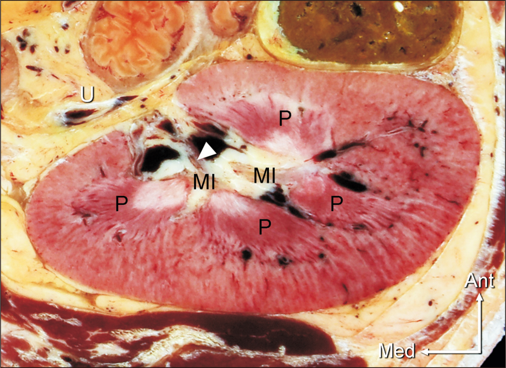
- Present case report describes a case of bifid ureter arising directly from separate calyces and renal pelvis of the kidney. Incomplete ureter duplication on the left side in a 78-year-old male cadaver was found during an anatomy class. These ureters converged in a Y-shaped pattern just above the level of the anterior superior iliac spine. In the coronal section of the kidney, the anterior ureter arose from a renal pelvis that was divided into two major calyces in the lower two-thirds of the kidney. On the other hand, the posterior ureter was directly connected to a major calyx in the upper third of the kidney, without the formation of a renal pelvis. This anatomical variation has implications for diagnostic approaches, especially in the use of imaging techniques by urologists for the insertion of stents in the treatment of phyelonephritis.
Keyword
Figure
Reference
-
References
1. Didier RA, Chow JS, Kwatra NS, Retik AB, Lebowitz RL. 2017; The duplicated collecting system of the urinary tract: embryology, imaging appearances and clinical considerations. Pediatr Radiol. 47:1526–38. DOI: 10.1007/s00247-017-3904-z. PMID: 29043421.2. Standring S. Gray's Anatomy: the anatomical basis of clinical practice. 42nd ed. Elsevier;2020.3. Tubbs RS, Shoja MM, Loukas M. Bergman's comprehensive encyclopedia of human anatomic variation. Wiley-Blackwell;2016. DOI: 10.1002/9781118430309.4. Privett JT, Jeans WD, Roylance J. 1976; The incidence and importance of renal duplication. Clin Radiol. 27:521–30. DOI: 10.1016/S0009-9260(76)80121-3. PMID: 1000896.5. Dorko F, Tokarčík J, Výborná E. 2016; Congenital malformations of the ureter: anatomical studies. Anat Sci Int. 91:290–4. DOI: 10.1007/s12565-015-0296-8. PMID: 26286110.6. Bhamani A, Srivastava M. 2013; Obstructed bifid uretereric system causing unilateral hydronephrosis. Rev Urol. 15:131–4. PMID: 24223026. PMCID: PMC3821993.7. Ravindra Swamy S, Kumar N, Patil J, Ashwini Aithal P, Guru A, Abhinitha P. 2014; Incomplete duplicated (bifid) left ureter - a case report. Sch J Med Case Rep. 2:303–5.8. Kumar V, Remya V. 2015; Unilateral incomplete bifid ureter: a case report. J Morphol Sci. 32:203–5. DOI: 10.4322/jms.077414.9. Ojha P, Prakash S. 2016; Unilateral incomplete duplicated ureter - a clinical and embryological insight. Int J Med Res Health Sci. 5:8:68–70.10. Shakthi Kumaran R, Chitra R. 2019; Unilateral duplex collecting system with incomplete duplication of ureters in right kidney in a male cadaver of Asian origin - a case report. Urol Case Rep. 23:99–100. DOI: 10.1016/j.eucr.2019.01.011. PMID: 30740309. PMCID: PMC6357695.11. Moinuddin Z, Dhanda R. 2015; Anatomy of the kidney and ureter. Anaesth Intensive Care Med. 16:247–52. DOI: 10.1016/j.mpaic.2015.04.001. PMID: 10685158.12. Anjana TS, Muthian E, Thiagarajan S, Shanmugam S. 2017; Gross morphological study of the renal pelvicalyceal patterns in human cadaveric kidneys. Indian J Urol. 33:36–40. DOI: 10.4103/0970-1591.194782. PMID: 28197028. PMCID: PMC5264190.13. Malligiannis Ntalianis K, Cheung C, Resta C, Liyanage S, Toneva F. 2022; Unilateral duplicated collecting system identified during pelvic lymphadenectomy: a case report and literature review. Cureus. 14:e26331. DOI: 10.7759/cureus.26331. PMID: 35775063. PMCID: PMC9236700.14. Protoshchak VV, Iglovikov NY, Shevnin MV. 2020; Combination of urolithiasis and anomaly: bifid ureter with fusion in the intramural part. Urol Ann. 12:196–8. DOI: 10.4103/UA.UA_142_19. PMID: 32565664. PMCID: PMC7292432.15. Sadler TW. Langman's medical embryology. 15th ed. Wolters Kluwer;2022.


