Anat Cell Biol.
2024 Sep;57(3):473-475. 10.5115/acb.23.289.
Complete transverse basilar cleft associated with hemifacial microsomia
- Affiliations
-
- 1Department of Anatomy, Faculty of Science, Mahidol University, Bangkok, Thailand
- 2In Silico and Clinical Anatomy Research Group (iSCAN), Bangkok, Thailand
- 3Department of Neurosurgery, Tulane Center for Clinical Neurosciences, Tulane University School of Medicine, New Orleans, LA, USA
- 4Department of Neurology, Tulane Center for Clinical Neurosciences, Tulane University School of Medicine, New Orleans, LA, USA
- 5Department of Oral and Maxillofacial Anatomy, Graduate School of Medical and Dental Sciences, Tokyo Medical and Dental University, Tokyo, Japan
- 6Department of Structural and Cellular Biology, Tulane University School of Medicine, New Orleans, LA, USA
- 7Department of Anatomy, Faculty of Medicine, Khon Kaen University, Khon Kaen, Thailand
- 8Department of Anatomy, Faculty of Medicine, Kasetsart University, Bangkok, Thailand
- 9Department of Diagnostic and Therapeutic Radiology, Faculty of Medicine Ramathibodi Hospital, Mahidol University, Bangkok, Thailand
- KMID: 2559769
- DOI: http://doi.org/10.5115/acb.23.289
Abstract
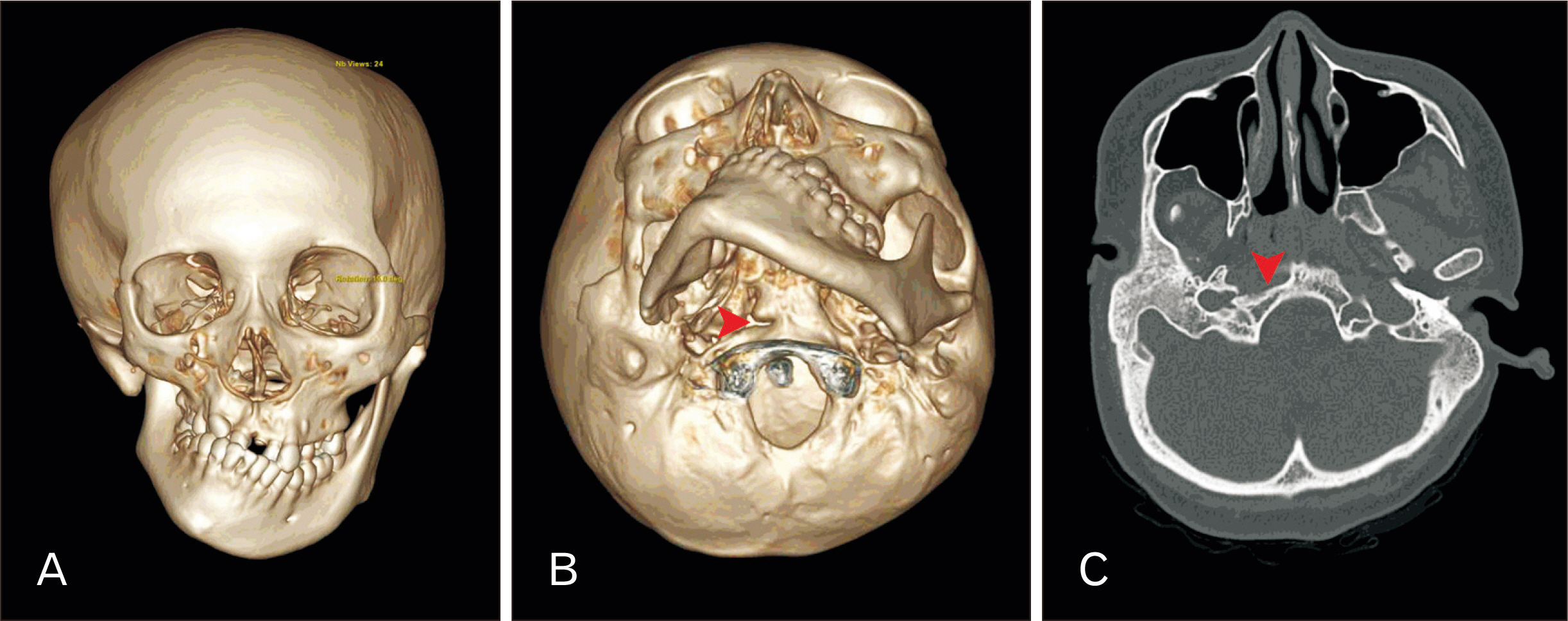
- Transverse basilar cleft (TBC) is an extremely rare variation of the clivus or the basilar part of the occipital bone. In this report, a unilateral transverse basilar fissure was found at the clivus in a head computed tomography of an 18-yearold female patient diagnosed with hemifacial microsomia (HFM). Image analysis of this patient showed shortening of the ramus of the right mandible along with medial displacement of the right temporomandibular joint and hypoplastic right maxilla. In addition, observation of the clivus showed a cleft between the basioticum and basioccipital bones at the level of the pharyngeal tubercle on the right side. This cleft was identified as TBC. Clival variations, TBC included, attributed to HFM have never been reported. This report draws attention to the complex relationship between abnormal development of clivus and HFM syndrome, and sheds light on a possible genetic and molecular association between these two conditions.
Figure
Reference
-
References
1. Hartsfield JK. 2007; Review of the etiologic heterogeneity of the oculo-auriculo-vertebral spectrum (Hemifacial microsomia). Orthod Craniofac Res. 10:121–8. DOI: 10.1111/j.1601-6343.2007.00391.x. PMID: 17651128.2. Murray JE, Kaban LB, Mulliken JB. 1984; Analysis and treatment of hemifacial microsomia. Plast Reconstr Surg. 74:186–99. DOI: 10.1097/00006534-198408000-00003. PMID: 6463144.3. Jinkins JR. Atlas of neuroradiologic embryology, anatomy, and variants. Lippincott Williams & Wilkins;2000.4. List CF. 1941; Neurologic syndromes accompanying developmental anomalies of occipital bone, atlas and axis. Arch Neurol Psychiatry. 45:577–616. DOI: 10.1001/archneurpsyc.1941.02280160009001.5. Barth J. Norrønaskaller: crania antiqua in parte orientali Norvegiae meridionalis inventa: en studie fra universitetets anatomiske institut. A.W. Brøggers bogtrykkeri;1896.6. Mahdi ES, Whitehead MT. 2018; Clival malformations in CHARGE syndrome. AJNR Am J Neuroradiol. 39:1153–6. DOI: 10.3174/ajnr.A5612. PMID: 29622552. PMCID: PMC7410616.7. Whitehead MT, Nagaraj UD, Pearl PL. 2015; Neuroimaging features of Cornelia de Lange syndrome. Pediatr Radiol. 45:1198–205. DOI: 10.1007/s00247-015-3300-5. PMID: 25701113.8. Tur SS, Svyatko SV, Rykun MP. 2019; Transverse basilar cleft: two more probable familial cases in an archaeological context. Int J Osteoarchaeol. 29:144–8. DOI: 10.1002/oa.2692.9. Köhler A, Zimmer EA, Schmidt H. Grenzen des normalen und anfänge des pathologischen im röntgenbild des skeletts. Thieme;1989.10. Lang J. Skull base and related structures: atlas of clinical anatomy. Schattauer;1995.11. Hofmann E, Prescher A. 2012; The clivus: anatomy, normal variants and imaging pathology. Clin Neuroradiol. 22:123–39. DOI: 10.1007/s00062-011-0083-4. PMID: 21710384.12. Psenner L. 1951; Die anatomischen varianten des hirnschädels. Röfo. 75:197–214. DOI: 10.1055/s-0029-1231984.13. Taiwo AO. 2020; Classification and management of hemifacial microsomia: a literature review. Ann Ib Postgrad Med. 18:S9–15. PMID: 33071690. PMCID: PMC7513375.14. Jensen BL. 1994; Cleidocranial dysplasia: craniofacial morphology in adult patients. J Craniofac Genet Dev Biol. 14:163–76. PMID: 7852545.15. Lou Y, Javed A, Hussain S, Colby J, Frederick D, Pratap J, Xie R, Gaur T, van Wijnen AJ, Jones SN, Stein GS, Lian JB, Stein JL. 2009; A Runx2 threshold for the cleidocranial dysplasia phenotype. Hum Mol Genet. 18:556–68. DOI: 10.1093/hmg/ddn383. PMID: 19028669. PMCID: PMC2638795.
- Full Text Links
- Actions
-
Cited
- CITED
-
- Close
- Share
- Similar articles
-
- Distraction osteogenesis in patients with hemifacial microsomia
- Clinical study of Simultaneous Correction of Bone and Soft Tissue Deformities in Hemifacial Microsmia
- Sequential treatment for a patient with hemifacial microsomia: 10 year-long term follow up
- A case report of hemifacial microsomia
- Three-dimensional functional unit analysis of hemifacial microsomia mandible-a preliminary report


