Anat Cell Biol.
2024 Sep;57(3):353-362. 10.5115/acb.23.316.
Prevalence of anatomical variants in the branches of celiac and superior mesenteric arteries among Egyptians
- Affiliations
-
- 1Department of Anatomy and Embryology, Faculty of Medicine, Tanta University, Tanta, Egypt
- 2Department of Basic and Clinical Oral Sciences, Faculty of Dental Medicine, Umm Al-Qura University, Makkah, Saudi Arabia
- KMID: 2559748
- DOI: http://doi.org/10.5115/acb.23.316
Abstract
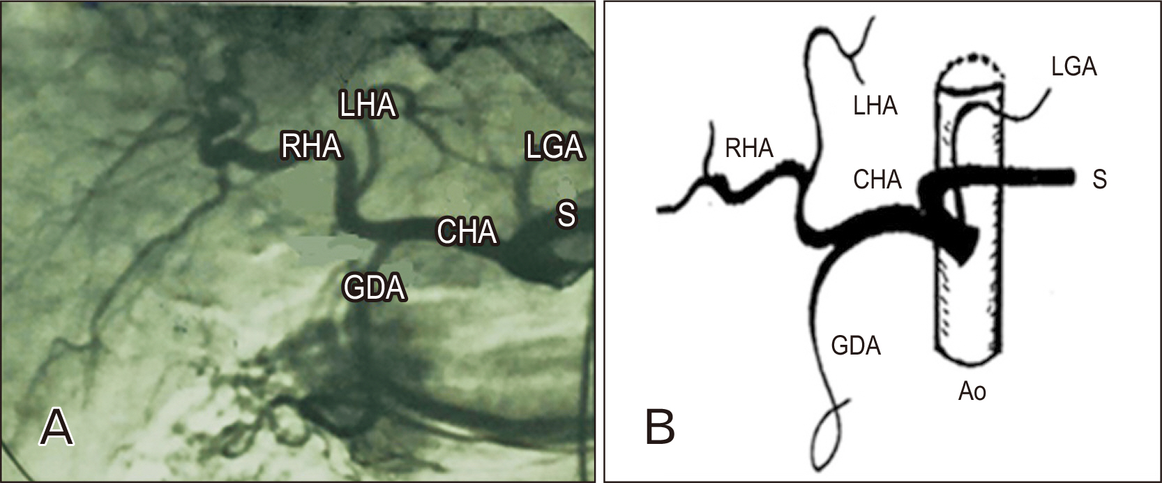
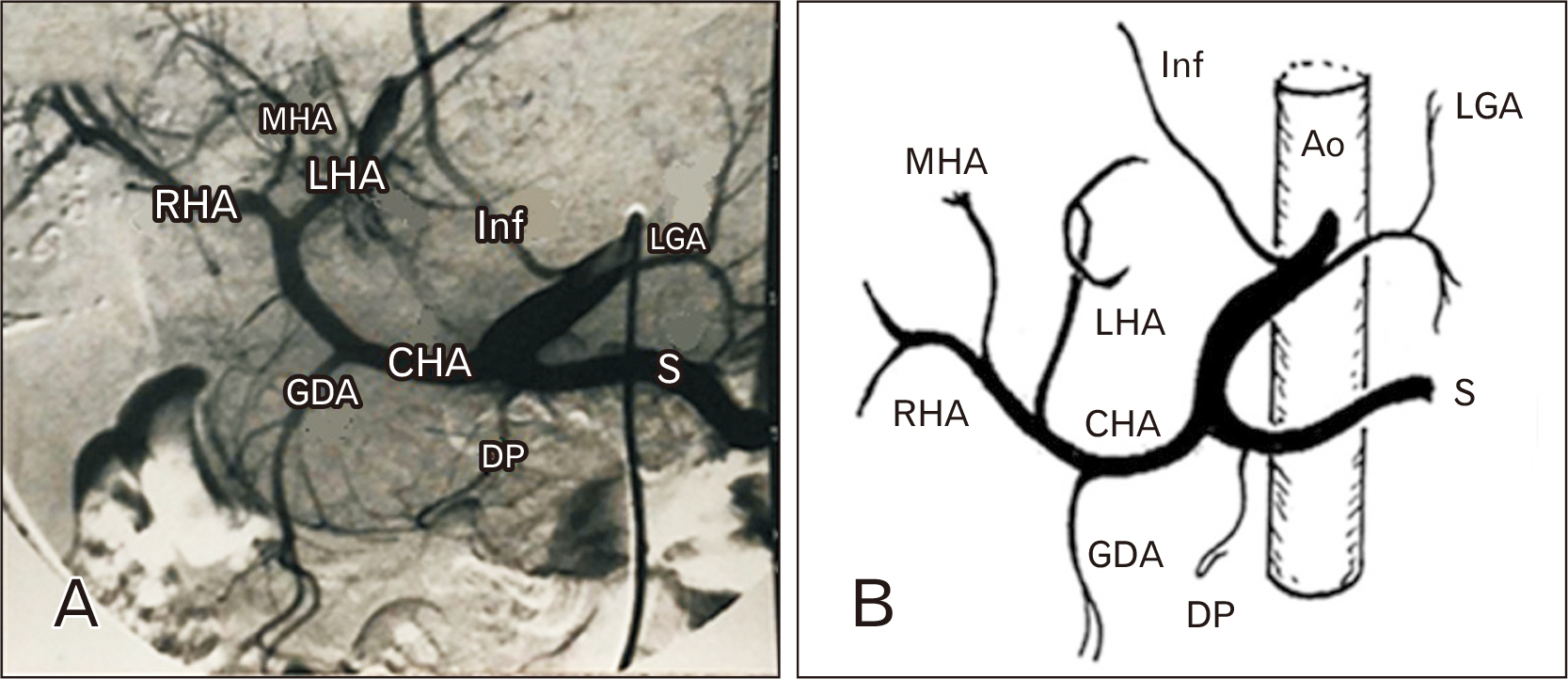
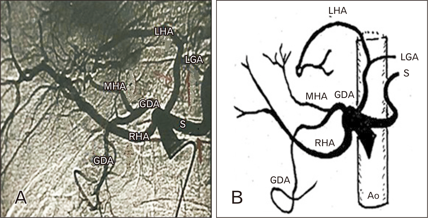
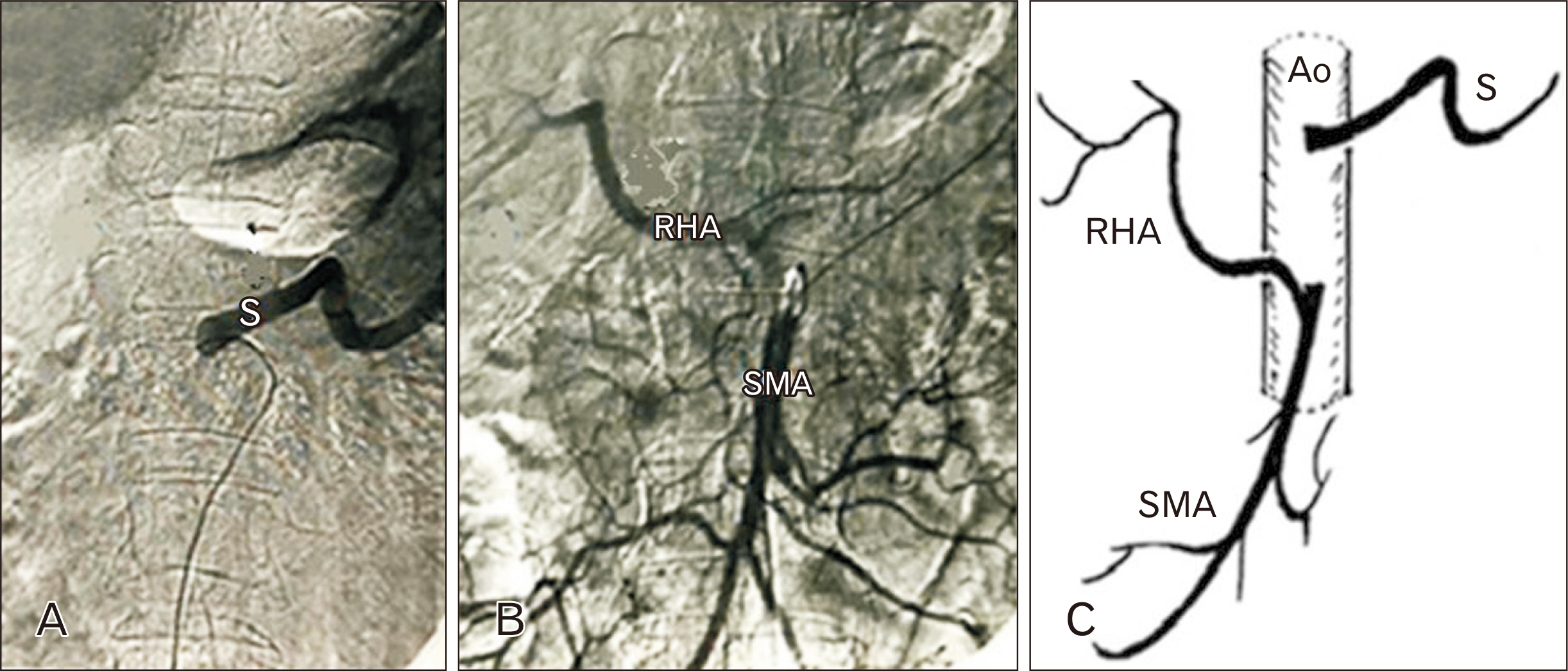
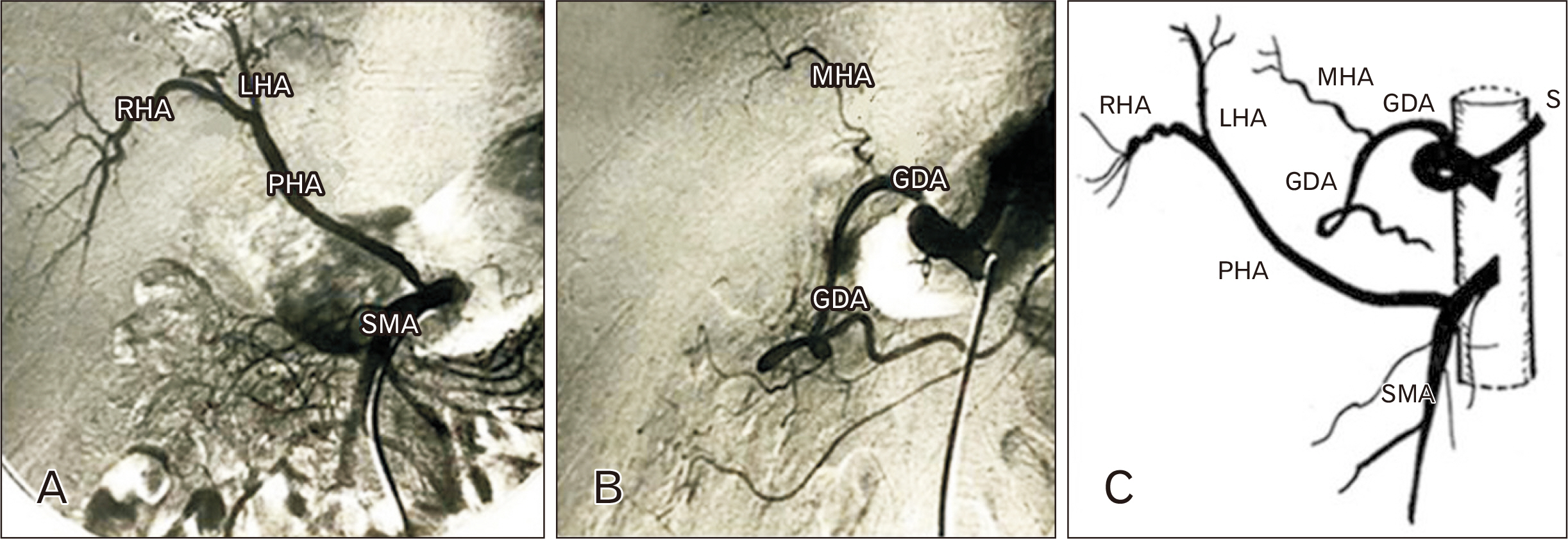
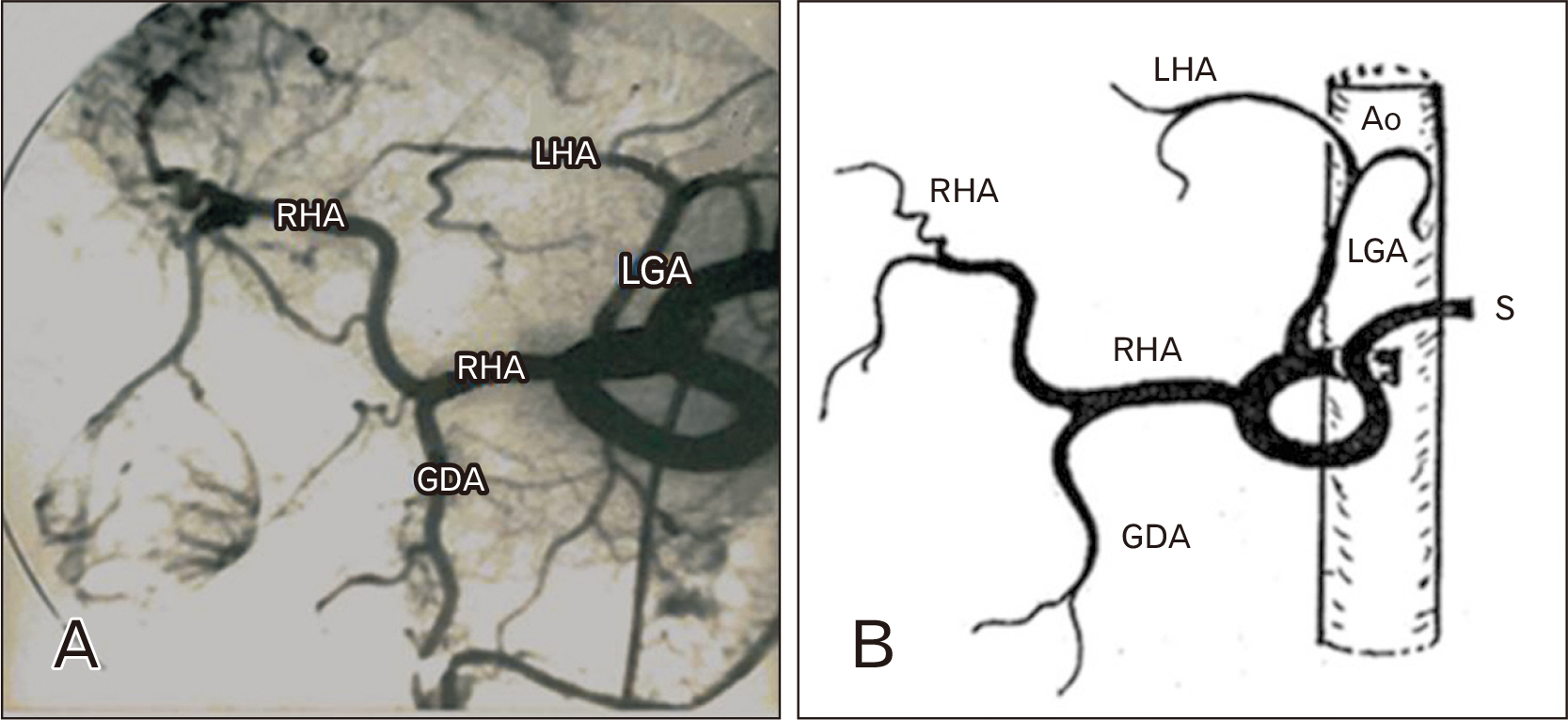
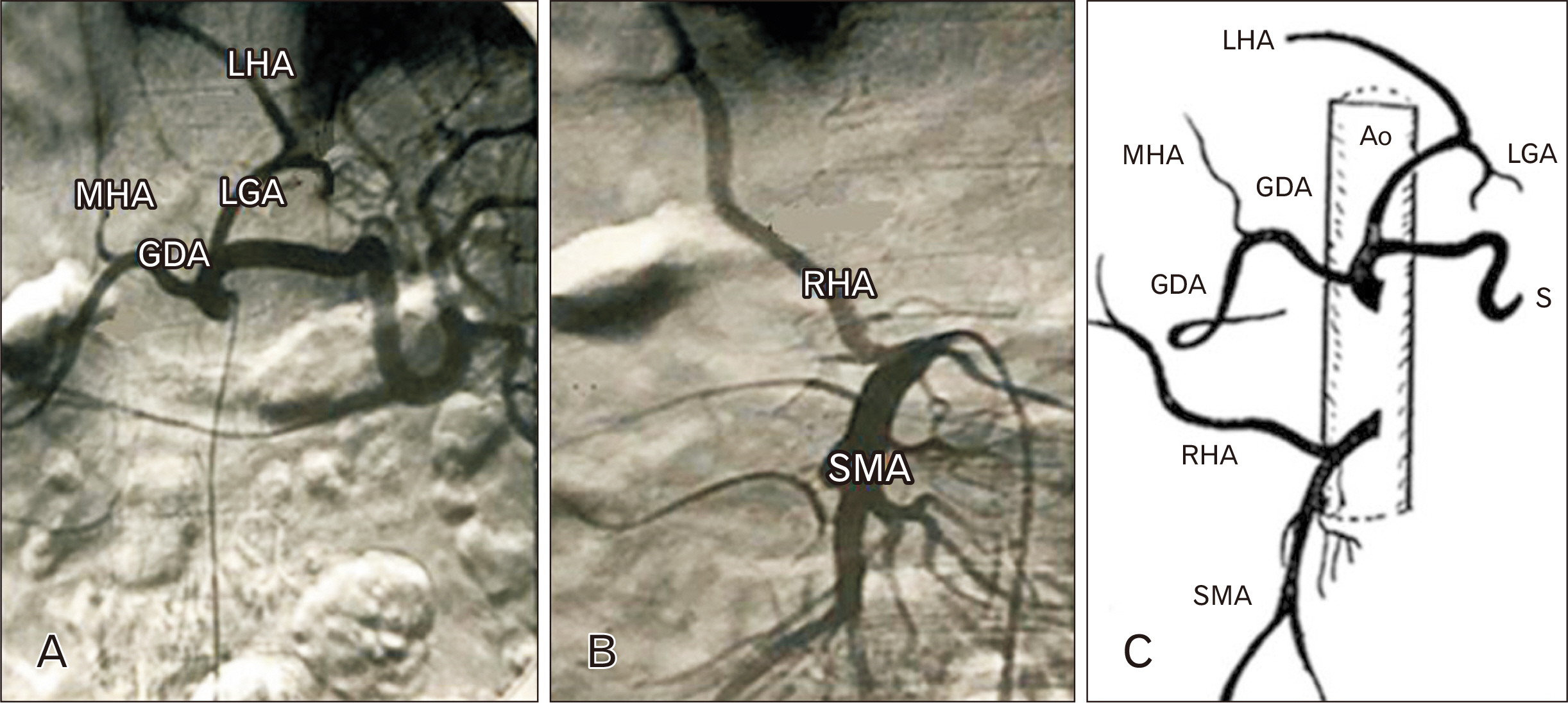
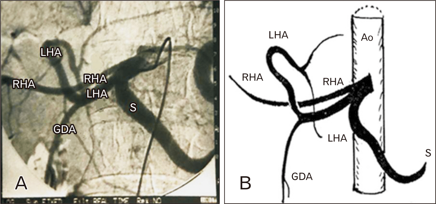
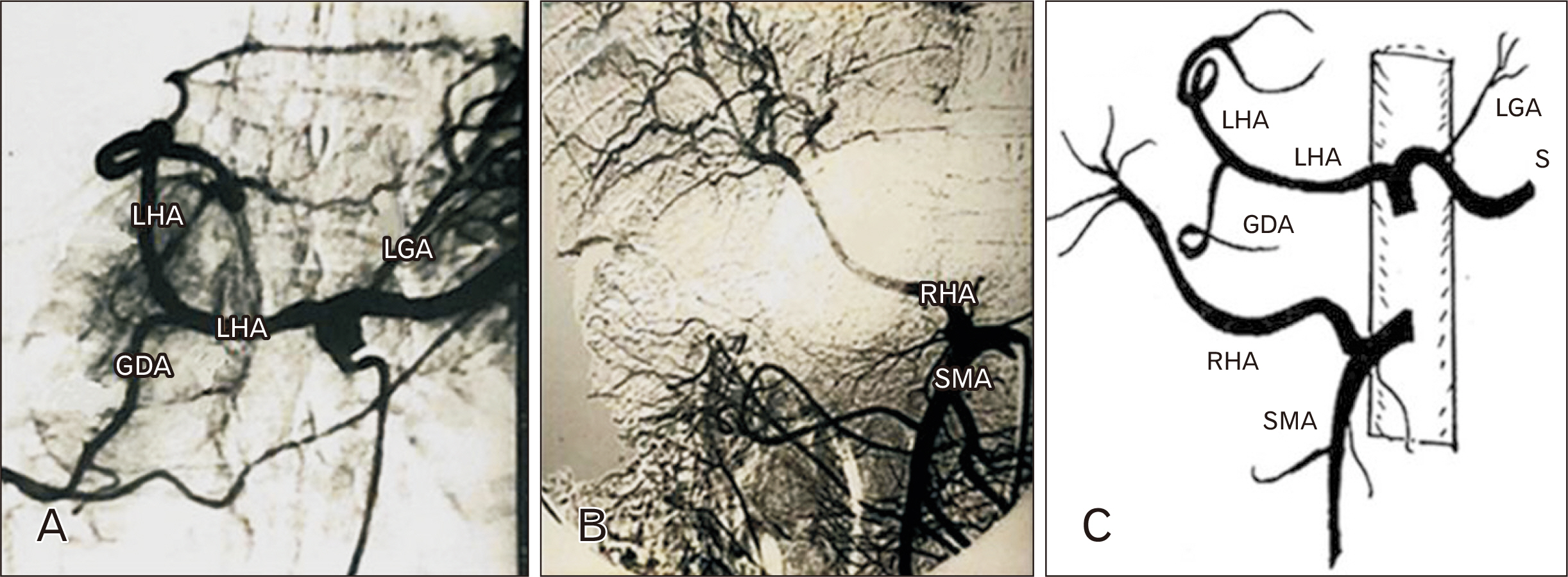
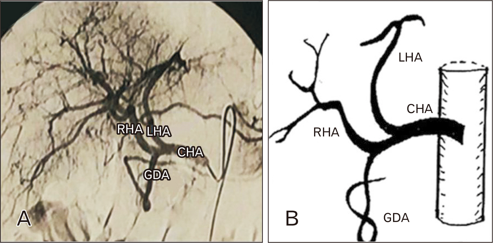
- Celiac trunk and superior mesenteric artery (SMA) are the main blood supply to the liver and pancreas. The data of anatomical variations in these arteries or their branches are very important clinically and surgically. The aim of this study was to describe the different variants in these arteries through the examination of the angiographs of a large series of Egyptian individuals. This research involved 389 selective angiographies to celiac artery, its branches, and the SMA. Anatomy of the target arteries of people who experienced visceral angiograph was reviewed and the data were recorded. From the total available angiograms in this work, 286 patients (73.52%) had the standard anatomy of celiac trunk and superior mesenteric arteries, and 103 patients (26.47%) had a single or multiple vessel variation. The inferior phrenic artery originates from celiac trunk in 2.05% of patients, while quadrifurcation of the celiac trunk was noticed in only 0.51% of patients. Absence of celiac trunk is also found in 0.51% of patients. Left gastric artery showed an abnormal origin from the splenic artery in 0.51% of patients. Quadrifurcation of common hepatic artery was also noticed. Variant anatomy of the left hepatic artery (LHA) was seen in 9.51% of patients, while variations of the right hepatic artery (RHA) were 14.13%. With the different origin of hepatic arteries, the gastroduodenal artery arose either from the LHA (2.82%), RHA (2.31%) or even from the celiac trunk (1.79%).
Figure
Reference
-
References
1. McMinn RMH. Last's anatomy: regional and applied. 8th ed. Churchill Livingstone;1990.2. Standring S. Gray's anatomy: the anatomical basis of clinical practice. 42nd ed. Elsevier;2020.3. Vandamme JP, Bonte J. 1985; The branches of the celiac trunk. Acta Anat (Basel). 122:110–4. DOI: 10.1159/000145991. PMID: 4013640.4. Noah EM, Klinzing S, Zwaan M, Schramm U, Bruch HP, Weiss HD. 1995; Normal variation of arterial liver supply in mesentericoceliacography. Ann Anat. 177:305–12. German. DOI: 10.1016/S0940-9602(11)80370-5. PMID: 7625603.5. Cavdar S, Gürbüz J, Zeybek A, Sehirli U, Abik L, Ozdogmuş O. 1998; A variation of coeliac trunk. Kaibogaku Zasshi. 73:505–8. PMID: 9844341.6. Covey AM, Brody LA, Maluccio MA, Getrajdman GI, Brown KT. 2002; Variant hepatic arterial anatomy revisited: digital subtraction angiography performed in 600 patients. Radiology. 224:542–7. DOI: 10.1148/radiol.2242011283. PMID: 12147854.7. Fasel JH, Muster M, Gailloud P, Mentha G, Terrier F. 1996; Duplicated hepatic artery: radiologic and surgical implications. Acta Anat (Basel). 157:164–8. DOI: 10.1159/000147878. PMID: 9142340.8. Gruttadauria S, Foglieni CS, Doria C, Luca A, Lauro A, Marino IR. 2001; The hepatic artery in liver transplantation and surgery: vascular anomalies in 701 cases. Clin Transplant. 15:359–63. DOI: 10.1034/j.1399-0012.2001.150510.x. PMID: 11678964.9. Marcos A, Orloff M, Mieles L, Olzinski A, Sitzmann J. 2001; Reconstruction of double hepatic arterial and portal venous branches for right-lobe living donor liver transplantation. Liver Transpl. 7:673–9. DOI: 10.1053/jlts.2001.26568. PMID: 11510010.10. Nakamura T, Tanaka K, Kiuchi T, Kasahara M, Oike F, Ueda M, Kaihara S, Egawa H, Ozden I, Kobayashi N, Uemoto S. 2002; Anatomical variations and surgical strategies in right lobe living donor liver transplantation: lessons from 120 cases. Transplantation. 73:1896–903. DOI: 10.1097/00007890-200206270-00008. PMID: 12131684.11. Jáuregui E. 1999; Anatomy of the splenic artery. Rev Fac Cien Med Univ Nac Cordoba. 56:21–41. Spanish. PMID: 10668264.12. Farghadani M, Momeni M, Hekmatnia A, Momeni F, Baradaran Mahdavi MM. 2016; Anatomical variation of celiac axis, superior mesenteric... artery, and hepatic artery: evaluation with multidetector computed tomography angiography. J Res Med Sci. 21:129. DOI: 10.4103/1735-1995.196611. PMID: 28331515. PMCID: PMC5348823.13. Santos PVD, Barbosa ABM, Targino VA, Silva NA, Silva YCM, Barbosa F, Oliveira ASB, Assis TO. 2018; Anatomical variations of the celiac trunk: a systematic review. Arq Bras Cir Dig. 31:e1403. DOI: 10.1590/0102-672020180001e1403. PMID: 30539978. PMCID: PMC6284376.14. Juszczak A, Mazurek A, Walocha JA, Pasternak A. 2021; Coeliac trunk and its anatomic variations: a cadaveric study. Folia Morphol (Warsz). 80:114–21. DOI: 10.5603/FM.a2020.0042. PMID: 32301103.15. Lippert H, Pabst R. Arterial variations in man: classification and frequency. J.F. Bergmann Verlag;1985.16. Wang Y, Cheng C, Wang L, Li R, Chen JH, Gong SG. 2014; Anatomical variations in the origins of the celiac axis and the superior mesenteric artery: MDCT angiographic findings and their probable embryological mechanisms. Eur Radiol. 24:1777–84. DOI: 10.1007/s00330-014-3215-9. PMID: 31197443.17. Michels NA. 1951; The hepatic, cystic and retroduodenal arteries and their relations to the biliary ducts with samples of the entire celiacal blood supply. Ann Surg. 133:503–24. DOI: 10.1097/00000658-195104000-00009. PMID: 14819988. PMCID: PMC1616853.18. Ghosh SK. 2014; Variations in the origin of middle hepatic artery: a cadaveric study and implications for living donor liver transplantation. Anat Cell Biol. 47:188–95. DOI: 10.5115/acb.2014.47.3.188. PMID: 25276478. PMCID: PMC4178194.19. Higashi N, Hirai K. 1995; A case of the three branches of the celiac trunk arising directly from the abdominal aorta. Kaibogaku Zasshi. 70:349–52. Japanese. PMID: 8540284.20. Başar R, Onderoğul S, Cumhur T, Yüksel M, Olçer T. 1995; Agenesis of the celiac trunk: an angiographic case. Kaibogaku Zasshi. 70:180–2. PMID: 7785416.21. Shoumura S, Emura S, Utsumi M, Chen H, Hayakawa D, Yamahira T, Isono H. 1991; Anatomical study on the branches of the celiac trunk (IV). Comparison of the findings with Adachi's classification. Kaibogaku Zasshi. 66:452–61. Japanese.22. Sponza M, Pozzi Mucelli R, Pozzi Mucelli F. 1993; Arterial anatomy of the celiac trunk and the superior mesenteric artery with computerized tomography. Radiol Med. 86:260–7. Italian. PMID: 8210535.23. Kadir S. Diagnostic angiography. Saunders;1986.24. Kahraman G, Marur T, Tanyeli E, Yildirim M. 2001; Hepatomesenteric trunk. Surg Radiol Anat. 23:433–5. DOI: 10.1007/s00276-001-0433-z. PMID: 11963627.25. Blumgart LH, Fong Y. Surgery of the liver and biliary tract. 3rd ed. W.B. Saunders;2000.26. Oran I, Yesildag A, Memis A. 2001; Aortic origin of right hepatic artery and superior mesenteric origin of splenic artery: two rare variations demonstrated angiographically. Surg Radiol Anat. 23:349–52. DOI: 10.1007/s00276-001-0349-7. PMID: 11824137.27. Sadler TW, Langman J. Langman's medical embryology. 6th ed. Williams and Wilkins;1990.
- Full Text Links
- Actions
-
Cited
- CITED
-
- Close
- Share
- Similar articles
-
- Accessory Renal Arteries Found during Dissection
- A rare variant angioarchitecture of upper abdomen
- Isolated Bypass to the Superior Mesenteric Artery for Chronic Mesenteric Ischemia
- A radiological study on normal variations of abdominal aorta and its major branches
- Dissecting Aneurysm of Superior Mesenteric Artery: A case report

