Obstet Gynecol Sci.
2024 Sep;67(5):506-510. 10.5468/ogs.24062.
Detection of trisomy 9 mosaicism in the second trimester screening by abnormal level of biochemical markers
- Affiliations
-
- 1Department of Obstetrics and Gynecology, School of Medicine, Neyshabur University of Medical Sciences, Neyshabur, Iran
- 2Department of Molecular Genetics, Faculty of Biological Sciences, Tarbiat Modares University, Tehran, Iran
- 3Healthy Ageing Research Center, School of Medicine, Neyshabur University of Medical Sciences, Neyshabur, Iran
- 4Department of Medical Biotechonology, School of Medicine, Neyshabur University of Medical Sciences, Neyshabur, Iran
- KMID: 2559482
- DOI: http://doi.org/10.5468/ogs.24062
Abstract
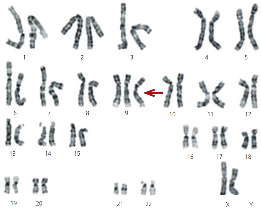
- Trisomy 9 is a rare chromosomal abnormality that occurs in both mosaic and non-mosaic states. The present study reports a case of mosaic trisomy 9 detected during pregnancy in a 41-year-old woman in the second trimester screening. Maternal serum screening results were used to diagnose a chromosomal abnormality in utero. The results were validated by karyotyping. High levels of alpha-fetoprotein and low levels of unconjugated estriol (uE3), human chorionic gonadotropin (hCG), and inhibin A indicate a high risk for chromosomal abnormalities, including trisomy 18. Amniotic fluid karyotyping revealed 47, XX, +9 (30)/46, XX (20) in the fetus. Because a high level (60%) of mosaicism for trisomy 9 in the fetus can affect many parts of the body, the pregnancy was terminated. It seems that a significant reduction in the levels of hCG and uE3 is an informative marker for the detection of chromosomal abnormalities such as trisomy 9.
Keyword
Figure
Reference
-
References
1. Feingold M, Atkins L. A case of trisomy 9. J Med Genet. 1973; 10:184–7.
Article2. Ma N, Zhu Z, Hu J, Pang J, Yang S, Liu J, et al. Case report: detection of fetal trisomy 9 mosaicism by multiple genetic testing methods: report of two cases. Front Genet. 2023; 14:1121121.
Article3. McDuffie RS Jr. Complete trisomy 9: case report with ultrasound findings. Am J Perinatol. 1994; 11:80–4.4. Chen CP, Chern SR, Cheng SJ, Chang TY, Yeh LF, Lee CC, et al. Second-trimester diagnosis of complete trisomy 9 associated with abnormal maternal serum screen results, open sacral spina bifida and congenital diaphragmatic hernia, and review of the literature. Prenat Diagn. 2004; 24:455–62.5. Bruns D. Presenting physical characteristics, medical conditions, and developmental status of long-term survivors with trisomy 9 mosaicism. Am J Med Genet A. 2011; 155A:1033–9.
Article6. Canick JA, MacRae AR. Second trimester serum markers. Semin Perinatol. 2005; 29:203–8.
Article7. Younesi S, Taheri Amin MM, Hantoushzadeh S, Saadati P, Jamali S, Modarressi MH, et al. Karyotype analysis of amniotic fluid cells and report of chromosomal abnormalities in 15,401 cases of Iranian women. Sci Rep. 2021; 11:19402.
Article8. Lam YH, Lee CP, Tang MH. Low second-trimester maternal serum human chorionic gonadotrophin in a trisomy 9 pregnancy. Prenat Diagn. 1998; 18:1212.9. Liu Y, Sun XC, Lv GJ, Liu JH, Sun C, Mu K. Amniotic fluid karyotype analysis and prenatal diagnosis strategy of 3117 pregnant women with amniocentesis indication. J Comp Eff Res. 2023; 12:e220168.
Article10. Lu S, Kakongoma N, Hu WS, Zhang YZ, Yang NN, Zhang W, et al. Detection rates of abnormalities in over 10,000 amniotic fluid samples at a single laboratory. BMC Pregnancy Childbirth. 2023; 23:102.
Article11. Hay SB, Sahoo T, Travis MK, Hovanes K, Dzidic N, Doherty C, et al. ACOG and SMFM guidelines for prenatal diagnosis: is karyotyping really sufficient? Prenat Diagn. 2018; 38:184–9.
Article12. Qian G, Cai L, Yao H, Dong X. Chromosome microarray analysis combined with karyotype analysis is a powerful tool for the detection in pregnant women with high-risk indicators. BMC Pregnancy Childbirth. 2023; 23:784.
Article13. Hao M, Li L, Zhang H, Li L, Liu R, Yu Y. The difference between karyotype analysis and chromosome microarray for mosaicism of aneuploid chromosomes in prenatal diagnosis. J Clin Lab Anal. 2020; 34:e23514.14. Stipoljev F, Kos M, Kos M, Miskovi B, Matijevic R, Hafner T, et al. Antenatal detection of mosaic trisomy 9 by ultrasound: a case report and literature review. J Matern Fetal Neonatal Med. 2003; 14:65–9.
Article15. Takahashi H, Hayashi S, Miura Y, Tsukamoto K, Kosaki R, Itoh Y, et al. Trisomy 9 mosaicism diagnosed in utero. Obstet Gynecol Int. 2010; 2010:379534.16. Chen CP, Lin HM, Su YN, Chern SR, Tsai FJ, Wu PC, et al. Mosaic trisomy 9 at amniocentesis: prenatal diagnosis and molecular genetic analyses. Taiwan J Obstet Gynecol. 2010; 49:341–50.
Article17. Chen CP, Hung FY, Su YN, Chern SR, Su JW, Lee CC, et al. Prenatal diagnosis of mosaic trisomy 9. Taiwan J Obstet Gynecol. 2011; 50:549–53.
Article18. Merino A, De Perdigo A, Nombalais F, Yvinec M, Le Roux MG, Bellec V. Prenatal diagnosis of trisomy 9 mosaicism: two new cases. Prenat Diagn. 1993; 13:1001–7.19. Zhang S, Zhou Y, Xiao G, Qiu X. Application of various genetic analysis techniques for detecting two rare cases of 9p duplication mosaicism during prenatal diagnosis. Mol Genet Genomic Med. 2023; 11:e2229.
Article


