J Liver Cancer.
2024 Sep;24(2):206-216. 10.17998/jlc.2024.05.01.
Re-assessing the diagnostic value of the enhancing capsule in hepatocellular carcinoma imaging
- Affiliations
-
- 1Department of Radiology, Seoul National University Hospital, Seoul, Korea
- 2Department of Radiology, Seoul National University College of Medicine, Seoul, Korea
- 3Institute of Radiation Medicine, Seoul National University Medical Research Center, Seoul, Korea
- 4Department of Radiology, Seoul National University Hospital Healthcare System Gangnam Center, Seoul, Korea
- 5G&E Alphadom Medical Center, Seongnam, Korea
- KMID: 2559467
- DOI: http://doi.org/10.17998/jlc.2024.05.01
Abstract
- Backgrounds/Aims
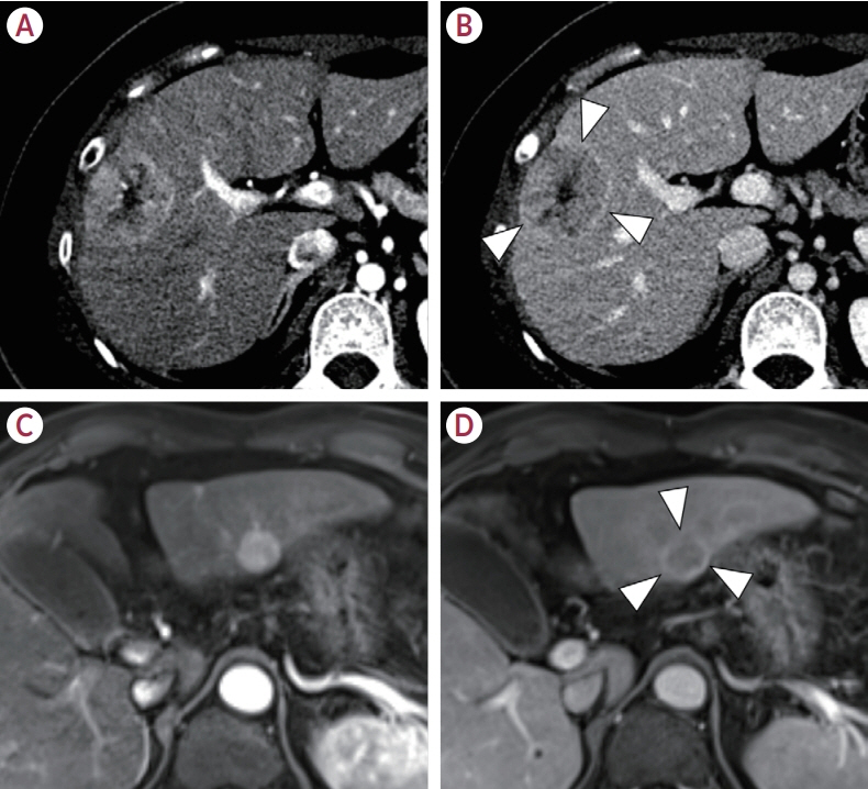
The enhancing capsule (EC) in hepatocellular carcinoma (HCC) diagnosis has received varying degrees of recognition across major guidelines. This study aimed to assess the diagnostic utility of EC in HCC detection.
Methods
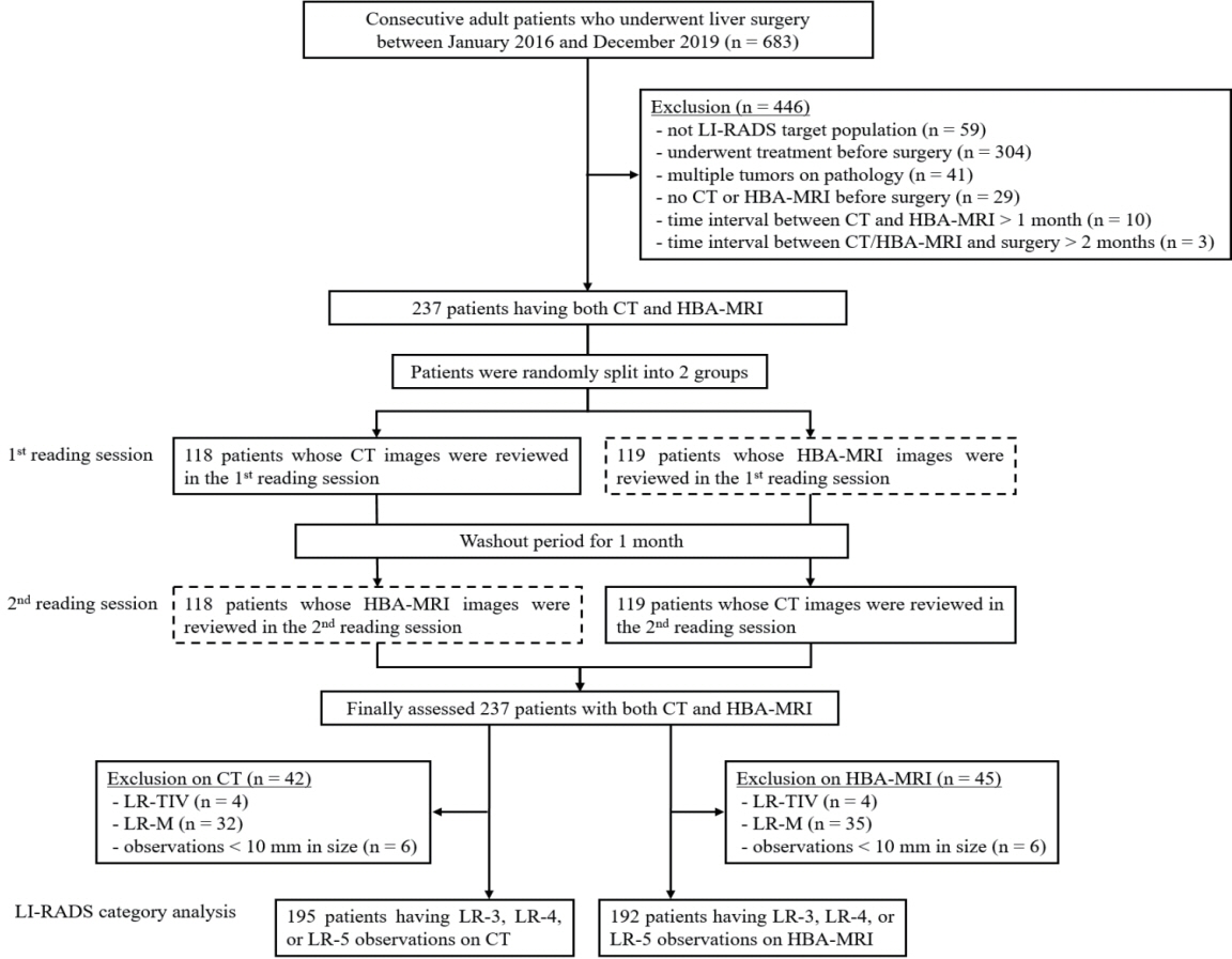
We retrospectively analyzed patients who underwent pre-surgical computed tomography (CT) and hepatobiliary agent-enhanced magnetic resonance imaging (HBA-MRI) between January 2016 and December 2019. A single hepatic tumor was confirmed based on the pathology of each patient. Three radiologists independently reviewed the images according to the Liver Imaging Reporting and Data System (LI-RADS) v2018 criteria and reached a consensus. Interobserver agreement for EC before reaching a consensus was quantified using Fleiss κ statistics. The impact of EC on the LI-RADS classification was assessed by comparing the positive predictive values for HCC detection in the presence and absence of EC.
Results
In total, 237 patients (median age, 60 years; 184 men) with 237 observations were included. The interobserver agreement for EC detection was notably low for CT (κ=0.169) and HBA-MRI (κ=0.138). The presence of EC did not significantly alter the positive predictive value for HCC detection in LI-RADS category 5 observations on CT (94.1% [80/85] vs. 94.6% [88/93], P=0.886) or HBAMRI (95.7% [88/92] vs. 90.6% [77/85], P=0.178).
Conclusions
The diagnostic value of EC in HCC diagnosis remains questionable, given its poor interobserver agreement and negligible impact on positive predictive values for HCC detection. This study challenges the emphasis on EC in certain diagnostic guidelines and suggests the need to re-evaluate its role in HCC imaging.
Figure
Reference
-
References
1. Llovet JM, Kelley RK, Villanueva A, Singal AG, Pikarsky E, Roayaie S, et al. Hepatocellular carcinoma. Nat Rev Dis Primers. 2021; 7:6.
Article2. Lee MW, Lee JM, Koh YH, Chung JW. 2022 Korean Liver Cancer Association-National Cancer Center Korea practice guidelines for local ablation therapy of hepatocellular carcinoma: what’s new? Korean J Radiol. 2023; 24:10–14.
Article3. Melendez-Torres J, Singal AG. Early detection of hepatocellular carcinoma: roadmap for improvement. Expert Rev Anticancer Ther. 2022; 22:621–632.
Article4. Kim JH, Joo I, Lee JM. Atypical appearance of hepatocellular carcinoma and its mimickers: how to solve challenging cases using gadoxetic acid-enhanced liver magnetic resonance imaging. Korean J Radiol. 2019; 20:1019–1041.5. Park J, Lee JM, Kim TH, Yoon JH. Imaging diagnosis of hepatocellular carcinoma: future directions with special emphasis on hepatobiliary magnetic resonance imaging and contrast-enhanced ultrasound. Clin Mol Hepatol. 2022; 28:362–379.
Article6. Chernyak V, Fowler KJ, Kamaya A, Kielar AZ, Elsayes KM, Bashir MR, et al. Liver Imaging Reporting and Data System (LI-RADS) version 2018: imaging of hepatocellular carcinoma in at-risk patients. Radiology. 2018; 289:816–830.
Article7. Consul N, Sirlin CB, Chernyak V, Fetzer DT, Masch WR, Arora SS, et al. Imaging features at the periphery: hemodynamics, pathophysiology, and effect on LI-RADS categorization. Radiographics. 2021; 41:1657–1675.
Article8. Shin J, Lee S, Yoon JK, Chung YE, Choi JY, Park MS. LI-RADS major features on MRI for diagnosing hepatocellular carcinoma: a systematic review and meta-analysis. J Magn Reson Imaging. 2021; 54:518–525.
Article9. van der Pol CB, McInnes MDF, Salameh JP, Levis B, Chernyak V, Sirlin CB, et al. CT/MRI and CEUS LI-RADS major features association with hepatocellular carcinoma: individual patient data meta-analysis. Radiology. 2022; 302:326–335.10. Razek AAKA, El-Serougy LG, Saleh GA, El-Wahab RA, Shabana W. Interobserver agreement of magnetic resonance imaging of Liver Imaging Reporting and Data System version 2018. J Comput Assist Tomogr. 2020; 44:118–123.
Article11. Kang JH, Choi SH, Lee JS, Kim KW, Kim SY, Lee SS, et al. Inter-reader reliability of CT Liver Imaging Reporting and Data System according to imaging analysis methodology: a systematic review and meta-analysis. Eur Radiol. 2021; 31:6856–6867.
Article12. Kang JH, Choi SH, Lee JS, Park SH, Kim KW, Kim SY, et al. Interreader agreement of Liver Imaging Reporting and Data System on MRI: a systematic review and meta-analysis. J Magn Reson Imaging. 2020; 52:795–804.
Article13. Rimola J, Sapena V, Brancatelli G, Darnell A, Forzenigo L, MähringerKunz A, et al. Reliability of extracellular contrast versus gadoxetic acid in assessing small liver lesions using Liver Imaging Reporting and Data System v.2018 and European Association for the Study of the Liver criteria. Hepatology. 2022; 76:1318–1328.
Article14. European Association for the Study of the Liver. EASL clinical practice guidelines: management of hepatocellular carcinoma. J Hepatol. 2018; 69:182–236.15. Korean Liver Cancer Association (KLCA); National Cancer Center (NCC) Korea. 2022 KLCA-NCC Korea practice guidelines for the management of hepatocellular carcinoma. Clin Mol Hepatol. 2022; 28:583–705.16. Korean Liver Cancer Association (KLCA); National Cancer Center (NCC) Korea. 2022 KLCA-NCC Korea practice guidelines for the management of hepatocellular carcinoma. J Liver Cancer. 2023; 23:1–120.17. Joo I, Lee JM, Koh YH, Choi SH, Lee S, Chung JW. 2022 Korean Liver Cancer Association-National Cancer Center Korea practice guidelines for imaging diagnosis of hepatocellular carcinoma: what’s new? Korean J Radiol. 2023; 24:1–5.18. Kang HJ, Lee JM, Jeon SK, Jang S, Park S, Joo I, et al. Intra-individual comparison of dual portal venous phases for non-invasive diagnosis of hepatocellular carcinoma at gadoxetic acid-enhanced liver MRI. Eur Radiol. 2021; 31:824–833.
Article19. American College of Radiology. Liver Imaging Reporting & Data System (LI-RADS®) [Internet]. Reston, VA (US): American College of Radiology;[cited 2024 Mar 8]. Available from: https://www.acr.org/ClinicalResources/Reporting-and-Data-Systems/LI-RADS.20. Pan J, Song M, Yang L, Zhao Y, Zhu Y, Wang M, et al. The role of enhancing capsule and modified capsule appearances in LI-RADS for diagnosing HCC ≤ 3.0 cm on gadoxetate disodium-enhanced MRI. Eur Radiol. 2023; 33:5801–5811.
Article21. Sofue K, Sirlin CB, Allen BC, Nelson RC, Berg CL, Bashir MR. How reader perception of capsule affects interpretation of washout in hypervascular liver nodules in patients at risk for hepatocellular carcinoma. J Magn Reson Imaging. 2016; 43:1337–1345.
Article22. Lee S, Kim SS, Roh YH, Choi JY, Park MS, Kim MJ. Diagnostic performance of CT/MRI Liver Imaging Reporting and Data System v2017 for hepatocellular carcinoma: a systematic review and meta-analysis. Liver Int. 2020; 40:1488–1497.
Article23. Burgio MD, Picone D, Cabibbo G, Midiri M, Lagalla R, Brancatelli G. MRimaging features of hepatocellular carcinoma capsule appearance in cirrhotic liver: comparison of gadoxetic acid and gadobenate dimeglumine. Abdom Radiol (NY). 2016; 41:1546–1554.24. Grazioli L, Olivetti L, Fugazzola C, Benetti A, Stanga C, Dettori E, et al. The pseudocapsule in hepatocellular carcinoma: correlation between dynamic MR imaging and pathology. Eur Radiol. 1999; 9:62–67.
Article25. Hope TA, Aslam R, Weinstein S, Yeh BM, Corvera CU, Monto A, et al. Change in Liver Imaging Reporting and Data System characterization of focal liver lesions using gadoxetate disodium magnetic resonance imaging compared with contrast-enhanced computed tomography. J Comput Assist Tomogr. 2017; 41:376–381.
Article26. Joo I, Lee JM, Lee DH, Ahn SJ, Lee ES, Han JK. Liver Imaging Reporting and Data System v2014 categorization of hepatocellular carcinoma on gadoxetic acid-enhanced MRI: comparison with multiphasic multidetector computed tomography. J Magn Reson Imaging. 2017; 45:731–740.
Article27. Kim B, Lee JH, Kim JK, Kim HJ, Kim YB, Lee D. The capsule appearance of hepatocellular carcinoma in gadoxetic acid-enhanced MR imaging: correlation with pathology and dynamic CT. Medicine (Baltimore). 2018; 97:e11142.28. Kim YN, Song JS, Moon WS, Hwang HP, Kim YK. Intra-individual comparison of hepatocellular carcinoma imaging features on contrast-enhanced computed tomography, gadopentetate dimeglumine-enhanced MRI, and gadoxetic acid-enhanced MRI. Acta Radiol. 2018; 59:639–648.29. Kim JH, Lee JY, Yu SJ, Lee DH, Joo I, Yoon JH, et al. Fusion imaging-guided radiofrequency ablation with artificial ascites or pleural effusion in patients with hepatocellular carcinomas: the feasibility rate and midterm outcome. Int J Hyperthermia. 2023; 40:2213424.
Article30. Lee S, Kim SS, Chang DR, Kim H, Kim MJ. Comparison of LI-RADS 2018 and KLCA-NCC 2018 for noninvasive diagnosis of hepatocellular carcinoma using magnetic resonance imaging. Clin Mol Hepatol. 2020; 26:340–351.
Article31. Lee SM, Lee JM, Ahn SJ, Kang HJ, Yang HK, Yoon JH. Diagnostic performance of 2018 KLCA-NCC practice guideline for hepatocellular carcinoma on gadoxetic acid-enhanced MRI in patients with chronic hepatitis B or cirrhosis: comparison with LI-RADS version 2018. Korean J Radiol. 2021; 22:1066–1076.
Article32. Park SH, Shim YS, Kim B, Kim SY, Kim YS, Huh J, et al. Retrospective analysis of current guidelines for hepatocellular carcinoma diagnosis on gadoxetic acid-enhanced MRI in at-risk patients. Eur Radiol. 2021; 31:4751–4763.
Article33. Choi SH, Jeon SK, Lee SS, Lee JM, Hur BY, Kang HJ, et al. Radiopathologic correlation of biphenotypic primary liver cancer (combined hepatocellular cholangiocarcinoma): changes in the 2019 WHO classification and impact on LI-RADS classification at liver MRI. Eur Radiol. 2021; 31:9479–9488.34. Choi SH, Lee SS, Park SH, Kim KM, Yu E, Park Y, et al. LI-RADS classification and prognosis of primary liver cancers at gadoxetic acid-enhanced MRI. Radiology. 2019; 290:388–397.
Article
- Full Text Links
- Actions
-
Cited
- CITED
-
- Close
- Share
- Similar articles
-
- Imaging Diagnosis of Hepatocellular Carcinoma
- Diagnosis of hepatocellular carcinoma: Which MRI contrast agent? Which diagnostic criteria?
- Recent development of diagnostic imaging of hepatocellular carcinoma
- Intraductal malignant tumors in the liver mimicking cholangiocarcinoma: Imaging features for differential diagnosis
- Surveillance of hepatocellular carcinoma: is only ultrasound enough?


