Clin Endosc.
2024 Sep;57(5):683-687. 10.5946/ce.2024.051.
A rare case of esophageal mucoepidermoid carcinoma successfully treated via endoscopic submucosal dissection
- Affiliations
-
- 1Department of Internal Medicine, Pusan National University School of Medicine and Biomedical Research Institute, Pusan National University Hospital, Busan, Korea
- 2Department of Pathology, Pusan National University School of Medicine and Biomedical Research Institute, Pusan National University Hospital, Busan, Korea
- KMID: 2559220
- DOI: http://doi.org/10.5946/ce.2024.051
Abstract
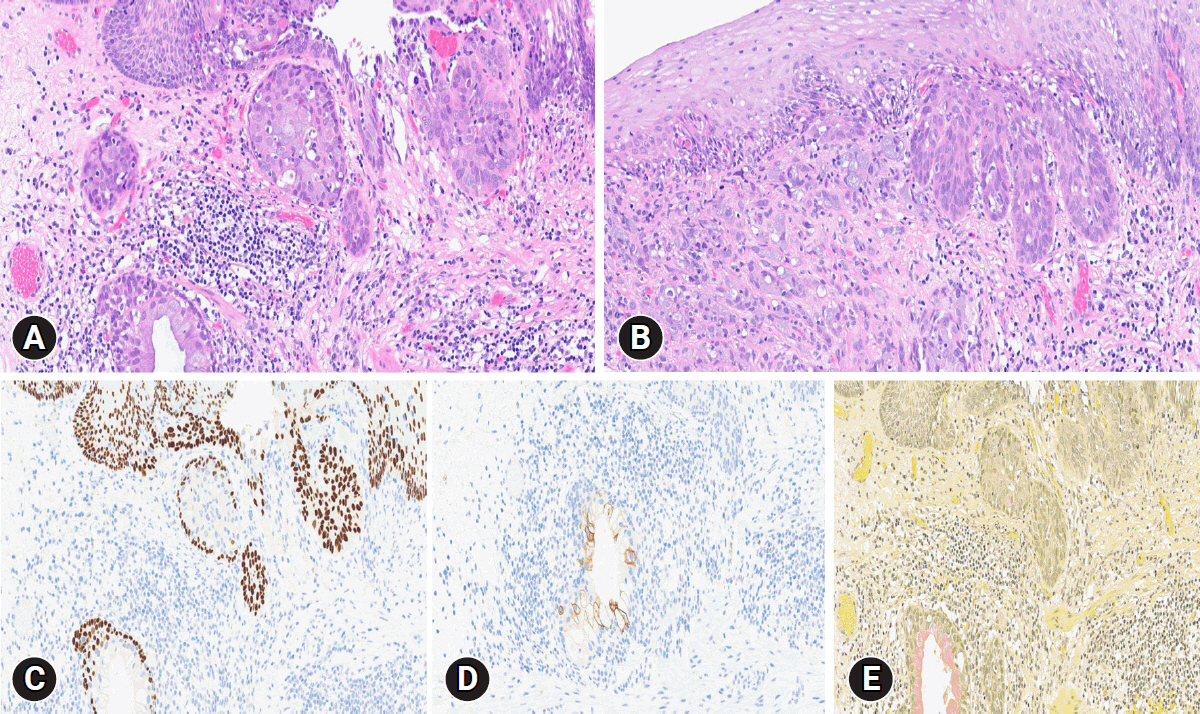
- Esophageal mucoepidermoid carcinoma (EMEC) is a special subtype of esophageal malignancy, accounting for less than 1% of all cases of primary esophageal carcinoma. Pathologically, it consists of a mixture of adenocarcinoma and squamous cell carcinoma with mucin-secreting cells. Special staining for mucicarmine helps to diagnose EMEC. We present a rare case of EMEC successfully treated via endoscopic submucosal dissection (ESD). A 63-year-old man was referred to our tertiary hospital. On esophagogastroduodenoscopy, a 6-mm-sized subtle reddish depressed lesion was identified in the mid-esophagus. Diagnostic ESD was performed with a high suspicion of carcinoma. Histopathologic findings were consistent with EMEC which was confined to the lamina propria without lymphatic invasion. We plan to do a careful follow-up without administering adjuvant chemotherapy or radiotherapy. Due to the small volume of the lesion, establishing a diagnosis was difficult through forceps biopsy alone. However, by using ESD, we could confirm and successfully treat a rare case of early-stage EMEC.
Keyword
Figure
Reference
-
1. Ferlay J, Soerjomataram I, Dikshit R, et al. Cancer incidence and mortality worldwide: sources, methods and major patterns in GLOBOCAN 2012. Int J Cancer. 2015; 136:E359–E386.2. Jung HK. Epidemiology of and risk factors for esophageal cancer in Korea. Korean J Helicobacter Up Gastrointest Res. 2019; 19:145–148.3. Abnet CC, Arnold M, Wei WQ. Epidemiology of esophageal squamous cell carcinoma. Gastroenterology. 2018; 154:360–373.4. Hagiwara N, Tajiri T, Tajiri T, et al. Biological behavior of mucoepidermoid carcinoma of the esophagus. J Nippon Med Sch. 2003; 70:401–407.5. Takubo K. Carcinomas other than squamous cell carcinoma and adenocarcinoma. In : Takubo K, editor. Pathology of the esophagus: an atlas and textbook. Springer;2007. p. 212–225.6. McPeak E, Arons WL. Adenoacanthoma of the esophagus: a report of one case with consideration of the tumor's resemblance to so-called salivary gland tumor. Arch Pathol (Chic). 1947; 44:385–390.7. Schizas D, Kapsampelis P, Mylonas KS. Adenosquamous carcinoma of the esophagus: a literature review. J Transl Int Med. 2018; 6:70–73.8. Liu ZJ, Sun SY, Guo JT, et al. A primary esophageal mucoepidermoid carcinoma mimicking a benign submucosal tumor. Dis Esophagus. 2012; 25:178–179.9. Tamura S, Kobayashi K, Seki Y, et al. Mucoepidermoid carcinoma of the esophagus treated by endoscopic mucosal resection. Dis Esophagus. 2003; 16:265–267.10. Stout AP, Latters RL. Tumor of the esophagus. In: Atlas of tumor pathology, Sect V, fasc 20. Armed Forces Institute of Pathology; 1957. p. 72–76.11. Kumagai Y, Ishiguro T, Kuwabara K, et al. Primary mucoepidermoid carcinoma of the esophagus: review of the literature. Esophagus. 2014; 11:81–88.12. Chen S, Chen Y, Yang J, et al. Primary mucoepidermoid carcinoma of the esophagus. J Thorac Oncol. 2011; 6:1426–1431.13. Koide N, Hamanaka K, Igarashi J, et al. Co-occurrence of mucoepidermoid carcinoma and squamous cell carcinoma of the esophagus: report of a case. Surg Today. 2000; 30:636–642.14. Wang X, Chen YP, Chen SB. Esophageal mucoepidermoid carcinoma: a review of 58 cases. Front Oncol. 2022; 12:836352.15. Suzuki Y, Nomura K, Matsui A, et al. Long-term outcomes of endoscopic submucosal dissection for special type of esophageal cancer. Dig Dis. 2023; 41:533–542.
- Full Text Links
- Actions
-
Cited
- CITED
-
- Close
- Share
- Similar articles
-
- Pyloric Gland Adenoma of the Esophagus Treated by Endoscopic Submucosal Dissection: A Case Report
- Perforation of a Gastric Tear during Esophageal Endoscopic Submucosal Dissection under General Anesthesia
- A Case of Esophageal Submucosal Dissection that Developed after Endoscopic Biopsy
- Superficial Esophageal Neoplasms Overlying Leiomyomas Removed by Endoscopic Submucosal Dissection: Case Reports and Review of the Literature
- Endoscopic Treatment for Esophageal Cancer


