Clin Exp Otorhinolaryngol.
2024 Aug;17(3):234-240. 10.21053/ceo.2024.00099.
Objective Parameters for Evaluating Internal Nasal Valve Compromise: Beyond the Angle Perspective
- Affiliations
-
- 1Department of Otorhinolaryngology-Head and Neck Surgery, Kyung Hee University Hospital at Gangdong, Kyung Hee University College of Medicine, Seoul, Korea
- KMID: 2558840
- DOI: http://doi.org/10.21053/ceo.2024.00099
Abstract
Objectives
. Nasal valve surgery for internal nasal valve (INV) compromise has become increasingly popular. However, this rise in popularity has sparked debates regarding its indications and disputes over insurance coverage, primarily due to the lack of a gold-standard evaluation method. Therefore, we aimed to identify objective parameters for the INV compromise.
Methods
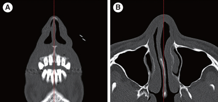
. We analyzed 186 INVs in 93 patients who underwent nasal valve surgery. The data comprised facial computed tomography (CT) images, acoustic rhinometry, the modified Cottle test, and symptom scores. Patients were categorized based on their symptoms and the results of the modified Cottle test. We measured the INV angle, area, volume, lateral wall thickness, septal angle, and nasal bone area using CT.
Results
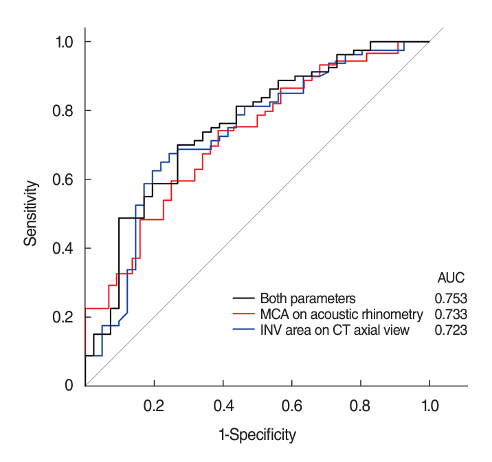
. The compromised INV group, characterized by nasal obstruction with a positive modified Cottle test, exhibited smaller INV areas in both coronal and axial views, reduced INV volume in the axial view, and a thinner lateral wall in the coronal view (all P<0.05). Acoustic rhinometry indicated a smaller minimal cross-sectional area and volume in the compromised INV group (both P<0.001). Regression analysis demonstrated significant associations between a compromised INV and reduced INV area on the axial view, as well as the minimal cross-sectional area measured by acoustic rhinometry.
Conclusion
. Relying solely on the INV angle in CT scans has limitations in assessing compromised INV. Alternatively, the INV area on axial CT scans and the minimal cross-sectional area measured by acoustic rhinometry may serve as objective parameters for evaluating INV compromise.
Keyword
Figure
Reference
-
1. Rhee JS, Weaver EM, Park SS, Baker SR, Hilger PA, Kriet JD, et al. Clinical consensus statement: diagnosis and management of nasal valve compromise. Otolaryngol Head Neck Surg. 2010; Jul. 143(1):48–59.2. Mink PJ. Le nez comme voie respiratoire. Presse Otolaryngol Belg. 1903; 21:481–96.3. Kasperbauer JL, Kern EB. Nasal valve physiology: implications in nasal surgery. Otolaryngol Clin North Am. 1987; Nov. 20(4):699–719.4. Suh MW, Jin HR, Kim JH. Computed tomography versus nasal endoscopy for the measurement of the internal nasal valve angle in Asians. Acta Otolaryngol. 2008; Jun. 128(6):675–9.5. Goudakos JK, Fishman JM, Patel K. A systematic review of the surgical techniques for the treatment of internal nasal valve collapse: where do we stand. Clin Otolaryngol. 2017; Feb. 42(1):60–70.6. Miman MC, Deliktaş H, Ozturan O, Toplu Y, Akarçay M. Internal nasal valve: revisited with objective facts. Otolaryngol Head Neck Surg. 2006; Jan. 134(1):41–7.7. San Nicolo M, Berghaus A, Jacobi C, Kisser U, Haack M, Flatz W. The nasal valve: new insights on the static and dynamic NV with MR-imaging. Eur Arch Otorhinolaryngol. 2020; Feb. 277(2):463–7.8. Cakmak O, Coskun M, Celik H, Buyuklu F, Ozluoglu LN. Value of acoustic rhinometry for measuring nasal valve area. Laryngoscope. 2003; Feb. 113(2):295–302.9. Kim SJ, Kim TH, Lee KH. An anatomic analysis of the bony vault: from the perspective of osteotomy in rhinoplasty. Aesthet Surg J. 2023; Apr. 43(5):535–42.10. Bloom JD, Sridharan S, Hagiwara M, Babb JS, White WM, Constantinides M. Reformatted computed tomography to assess the internal nasal valve and association with physical examination. Arch Facial Plast Surg. 2012; Sep-Oct. 14(5):331–5.11. Poetker DM, Rhee JS, Mocan BO, Michel MA. Computed tomography technique for evaluation of the nasal valve. Arch Facial Plast Surg. 2004; Jul-Aug. 6(4):240–3.12. Abdelwahab M, Yoon A, Okland T, Poomkonsarn S, Gouveia C, Liu SY. Impact of distraction osteogenesis maxillary expansion on the internal nasal valve in obstructive sleep apnea. Otolaryngol Head Neck Surg. 2019; Aug. 161(2):362–7.13. Moche JA, Cohen JC, Pearlman SJ. Axial computed tomography evaluation of the internal nasal valve correlates with clinical valve narrowing and patient complaint. Int Forum Allergy Rhinol. 2013; Jul. 3(7):592–7.14. Kim YM, Rha KS, Weissman JD, Hwang PH, Most SP. Correlation of asymmetric facial growth with deviated nasal septum. Laryngoscope. 2011; Jun. 121(6):1144–8.15. Ozturan O, Miman MC, Kizilay A. Bending of the upper lateral cartilages for nasal valve collapse. Arch Facial Plast Surg. 2002; Oct-Dec. 4(4):258–61.16. Yeung A, Hassouneh B, Kim DW. Outcome of nasal valve obstruction after functional and aesthetic-functional rhinoplasty. JAMA Facial Plast Surg. 2016; Mar-Apr. 18(2):128–34.17. Shafik AG, Alkady HA, Tawfik GM, Mohamed AM, Rabie TM, Huy NT. Computed tomography evaluation of internal nasal valve angle and area and its correlation with NOSE scale for symptomatic improvement in rhinoplasty. Braz J Otorhinolaryngol. 2020; May-Jun. 86(3):343–50.18. Friedman O, Cook TA. Conchal cartilage butterfly graft in primary functional rhinoplasty. Laryngoscope. 2009; Feb. 119(2):255–62.19. Bae MR, Choi WR, Jang YJ. The relationship between lateral nasal wall collapse and nasal obstruction. Ann Otol Rhinol Laryngol. 2023; Jul. 132(7):745–51.20. Ganjaei KG, Soler ZM, Mappus ED, Worley ML, Rowan NR, Garcia GJM, et al. Radiologic changes in the aging nasal cavity. Rhinology. 2019; Apr. 57(2):117–24.21. Kim SG, Menapace DC, Mims MM, Shockley WW, Clark JM. Age-related histologic and biochemical changes in auricular and nasal cartilages. Laryngoscope. 2024; Mar. 134(3):1220–6.22. Wiederkehr I, Kawabata Y, Tsumiyama S, Hosokawa Y, Iimura J, Otori N, et al. Caudal septal deviation: a computed tomography-based evaluation method. Ann Plast Surg. 2022; Jul. 89(1):95–9.23. Avashia YJ, Glener AD, Marcus JR. Functional nasal surgery. Plast Reconstr Surg. 2022; Aug. 150(2):439e–454e.
- Full Text Links
- Actions
-
Cited
- CITED
-
- Close
- Share
- Similar articles
-
- Surgery for Nasal Valve Compromise
- Measurement of the so-called "Nasal Valve" in Japanese Subjects
- How to Adjust the Thickness of the Spreader Graft in Nasal Valve Collapse
- Definition of Nasal Valve Location and Classification of Nasal Valve Stenosis in Computed Tomography
- Surgical Correction of Dynamic Nasal Valve Collapse


