Jejunal Dieulafoy’s lesion resembling subepithelial mass resulting in profound gastrointestinal hemorrhage
- Affiliations
-
- 1Division of Gastroenterology, Department of Medicine, Siriraj Hospital, Mahidol University, Bangkok, Thailand
- 2Department of Pathology, Siriraj Hospital, Mahidol University, Bangkok, Thailand
- 3Department of Radiology, Siriraj Hospital, Mahidol University, Bangkok, Thailand
- KMID: 2558115
- DOI: http://doi.org/10.5946/ce.2023.231
Figure
-
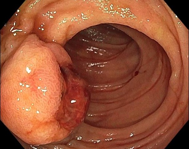
Fig. 1. Push enteroscopy reveals a 2.5 cm round subepithelial mass with a large central defect covered by an adherent clot in the proximal part of the jejunum.
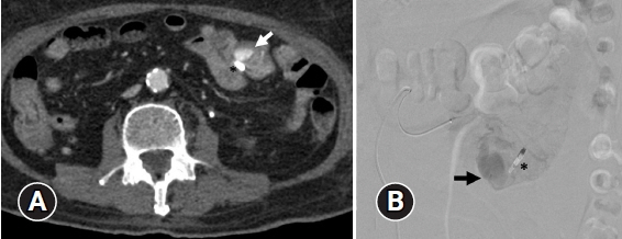
Fig. 2. (A) Arterial phase computed tomographic angiography shows a round-shaped 2×1.4 cm arterial enhancing lesion in the proximal jejunum (arrow) adjacent to hemostatic clip (*) without active contrast extravasation within bowel loops. (B) The angiography of the jejunal branch of the superior mesenteric artery reveals an accumulation of contrast media at the proximal jejunum (arrow) near the hemostatic clip (*), which is supplied by multiple small jejunal branches. No demonstrable active contrast extravasation is seen in the study.
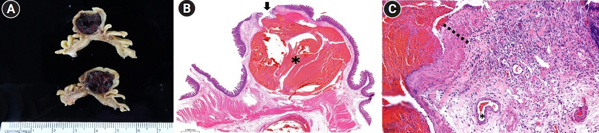
Fig. 3. (A) The gross lesion reveals a large thrombus within the submucosa. (B) The histopathology reveals a submucosal persistent-caliber artery, dilated with a recent thrombus (*) (hematoxylin and eosin stain, ×10). The overlying mucosal defect (arrow) is connected to the lesion. (C) The vascular wall of the lesion (dotted line) is exceedingly thickened compared to the normal submucosal artery (*) (hematoxylin and eosin stain, ×200).
Reference
-
1. Kim SE, Kim HJ, Koh M, et al. A practical approach for small bowel bleeding. Clin Endosc. 2023; 56:283–289.
Article2. Baxter M, Aly EH. Dieulafoy's lesion: current trends in diagnosis and management. Ann R Coll Surg Engl. 2010; 92:548–554.3. Malik TF, Anjum F. Dieulafoys Lesion causing gastrointestinal bleeding. StatPearls;2023.4. Sakai E, Ohata K, Nakajima A, et al. Diagnosis and therapeutic strategies for small bowel vascular lesions. World J Gastroenterol. 2019; 25:2720–2733.
Article5. Tahir MM, Saeed S, Hlaing SS, et al. Dieulafoy lesion causing lower GI bleeding: a case of COVID-19 critical illness prompting unusual presentation of a more rare condition. Critical Care. 2023; 162:A926–A927.
Article6. Malik A, Inayat F, Goraya MH, et al. Jejunal Dieulafoy's lesion: a systematic review of evaluation, diagnosis, and management. J Investig Med High Impact Case Rep. 2021; 9:2324709620987703.
Article7. Badwal S, Jain M, Rastogi A, et al. Dieulafoy disease of the stomach presenting as mass lesion: a case report. Indian J Pathol Microbiol. 2005; 48:211–213.8. Jeong HW, Kim JY, Kim SJ, et al. A case of a Jejunal Dieulafoy's lesion mimicking a submucosal tumor. Korean J Gastrointest Endosc. 2008; 37:438–442.9. Zhao J, Sun Z, Zhang X. A Jejunal Dieulafoy's lesion mimicking a gastrointestinal stromal tumor. Clin Gastroenterol Hepatol. 2019; 17:A19.
Article10. Shrestha S, Pradhan S, Kc A, et al. Arteriovenous malformation of the Jejunum, causing massive gastrointestinal bleeding, treated with intraoperative enteroscopy guidance: a case report. Cureus. 2023; 15:e39940.
Article
- Full Text Links
-
- Actions
-
Cited
- CITED
-
- Close
- Share
-
- Similar articles
-
- A Case of a Jejunal Dieulafoy's Lesion Mimicking a Submucosal Tumor
- A Case of Dieulafoy's Lesion in the Jejunum Treated by Double Balloon Enteroscopy
- Dieulafoy's Lesion of Jejunum: Presenting Small Bowel Mass and Stricture
- A Case of Dieulafoy Lesion of the Jejunum Presented with Massive Hemorrhage
- A case of bleeding from the Dieulafoy lesion of the jejunum
