Clin Endosc.
2024 Jul;57(4):547-548. 10.5946/ce.2024.037.
Closure of esophageal–pleural fistula using a cardiac occluder in a patient with systemic scleroderma
- Kiosov O
 1,3
1,3 - Tkachov V2,3
- Gulevskyi S3
- Affiliations
-
- 1Department of General Surgery and Postgraduate Surgical Education, Zaporizhzhia State Medical and Pharmaceutical University, Zaporizhzhia, Ukraine
- 2Department of Faculty Surgery, Zaporizhzhia State Medical and Pharmaceutical University, Zaporizhzhia, Ukraine
- 3Multidisciplinary Surgical Department, University Clinic of Zaporizhzhia State Medical and Pharmaceutical University, Zaporizhzhia, Ukraine
- KMID: 2558113
- DOI: http://doi.org/10.5946/ce.2024.037
Figure
-
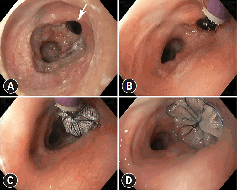
Fig. 1. (A) An image of the large 8 mm esophageal–pleural fistula (arrow) at 5 days before occluder placement. (B) Initiation of opening the occluder from the delivery device. (C) Detachment of the occluder from the delivery device. (D) Successful placement of the atrial septal defect occluder.
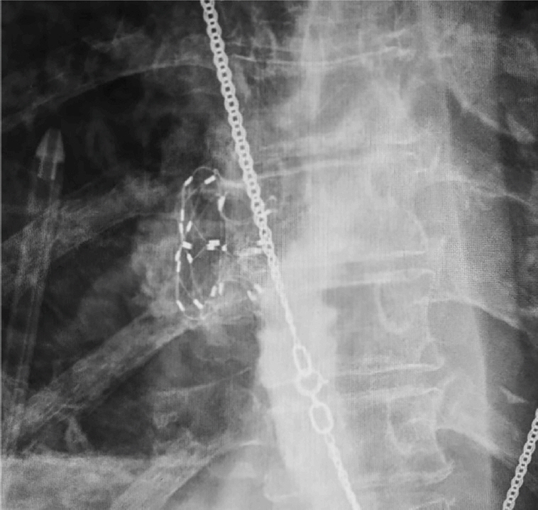
Fig. 2. X-ray taken the day after occluder placement.
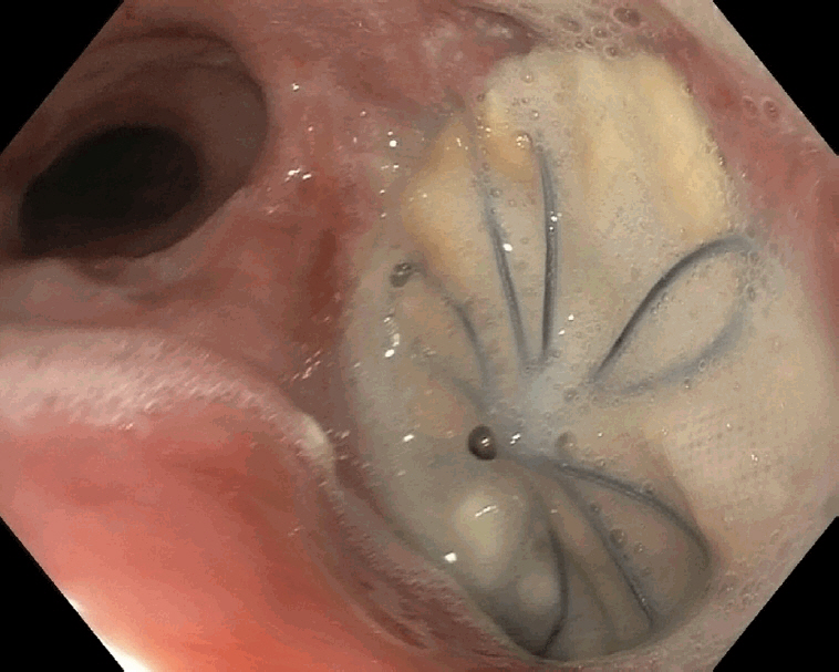
Fig. 3. Follow-up endoscopy performed 1 month after occluder placement.
Reference
-
1. Lassaletta AD, Laham RJ, Pinto DS, et al. Successful closure of an esophagopleural fistula with an amplatzer occluder sealed with liquid copolymer, with 3-year follow-up and review of literature. Pleura. 2016; 3:1–6.
Article2. Rosendahl AH, Schönborn K, Krieg T. Pathophysiology of systemic sclerosis (scleroderma). Kaohsiung J Med Sci. 2022; 38:187–195.
Article
- Full Text Links
-
- Actions
-
Cited
- CITED
-
- Close
- Share
-
- Similar articles
-
- The Management of Delayed Post-Pneumonectomy Broncho-Pleural Fistula and Esophago-Pleural Fistula
- A Case of Pleural Effusion due to Vasculitis in Scleroderma
- A Case Demonstrating a Percutaneous Closure Using the Amplatzer Duct Occluder for Paravalvular Leakage after Tricuspid Valve Replacement
- Late Migration of Amplatzer Septal Occluder Device to the Descending Thoracic Aorta
- Percutaneous Closure of the Acquired Gerbode Shunt Using the Amplatzer Duct Occluder in a 3-Month Old Patient
