Korean Circ J.
2024 Jul;54(7):425-426. 10.4070/kcj.2024.0095.
Surprising Course of a Pregnant Patient With Mosaic Turner Syndrome
- Affiliations
-
- 1Department of Cardiovascular Medicine, Mayo Clinic, Scottsdale, AZ, USA
- 2Department of Obstetrics, Gynecology and Women’s Health Institute, Cleveland Clinic, Cleveland, OH, USA
- 3Department of Obstetrics and Gynecology, Cleveland Clinic, Cleveland, OH, USA
- 4Department of Thoracic and Cardiovascular Surgery, Cleveland Clinic, Cleveland, OH, USA
- KMID: 2557983
- DOI: http://doi.org/10.4070/kcj.2024.0095
Figure
-
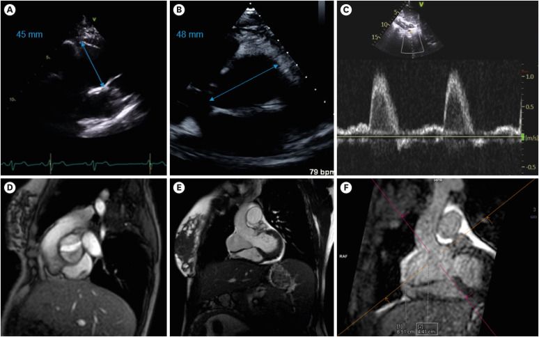
Figure 1 Select cardiac images of a pregnant patient with mosaic Turner syndrome. Echocardiographic parasternal long axis images revealing dilated aortic root at 45 mm (A), parasternal short axis images revealing an eccentrically dilated aortic root at 48 mm (B) and subcostal images revealing normal abdominal aortic Doppler pattern suggesting no residual coarctation (C). Magnetic resonance imaging revealing the bicuspid aortic valve (D) and eccentric aortic root dilation measuring 65 mm (E, F).
Reference
-
1. Shahrokhi Sabzevar S, Mirzaei F, Tanipour MH, Eslahi A, Hasanzadeh Nazarabadi M. Pregnancy in a patient with mosaic turner syndrome: a case report. CRCP. 2020; 5:58–62.
Article2. Yousif A, Wheatley M, Abuzeid MA. A case report of spontaneous pregnancy in a mosaic turner syndrome patient. Obstet Gynecol Cases Rev. 2020; 7:181.
Article
- Full Text Links
-
- Actions
-
Cited
- CITED
-
- Close
- Share
-
- Similar articles
-
- Mosaic Turner syndrome associated with schizophrenia
- A spontaneous pregnancy and Cesarean delivery in a Turner mosaic with previous recurrent miscarriages
- Ovarian mixed germ cell tumor in a patient with 45,X/46,X,+mar mosaic Turner's syndrome
- 45,X / 47,XYY Mosaic Turner Syndrome
- A case of 45,X/47,XXX mosaic Turner syndrome: Clinical manifestations and effect of growth hormone treatment
