Can Acanthamoeba keratitis be properly diagnosed without culture in the real-world clinical microbiology laboratory?: a case report
- Affiliations
-
- 1Department of Laboratory Medicine, Asan Medical Center, University of Ulsan College of Medicine, Seoul, Korea
- 2Department of Ophthalmology, Asan Medical Center, University of Ulsan College of Medicine, Seoul, Korea
- KMID: 2557875
- DOI: http://doi.org/10.5145/ACM.2024.27.2.9
Abstract
- Acanthamoeba species are ubiquitous, free-living organisms found in the environment. They can cause a sight-threatening cornea disease, termed Acanthamoeba keratitis, and are often misdiagnosed, causing delayed administration of the correct treatment. Herein, we report a case of Acanthamoeba keratitis diagnosed without culture. A 12-year-old girl with a history of wearing contact lenses presented with complaints of pain, irritation, and hyperemia in the left eye. Corneal scraping-smeared slide, and liquids with contact lenses were submitted to the clinical microbiology laboratory. Cultures of Acanthamoeba spp. were not available; thus, they were stained with calcofluor white. The isolation of Acanthamoeba from the corneal scraping allowed the detection of trophozoites and cysts based on their morphological characteristics. PCR targeting the 18s rRNA gene and subsequent sequencing revealed 99% identity with the Acanthamoeba spp. Although it is challenging to find real-world evidence of Acanthamoeba in clinical microbiology without using culture methods, this case underscores the need for clinical microbiology laboratories to maintain their inspection capabilities.
Keyword
Figure
-
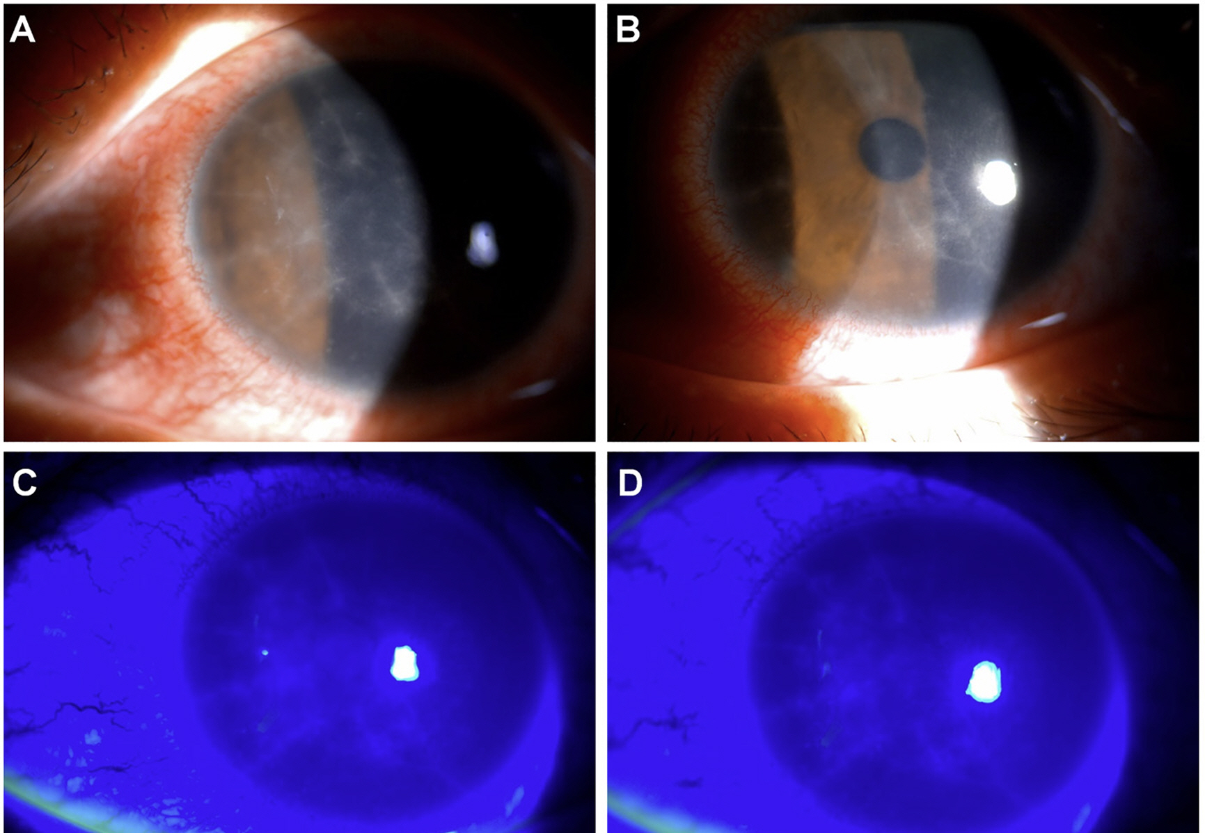
Fig. 1. Anterior segment photographs of the patient in this case study. The top two anterior segment images were taken under slit-lamp microscope (A, B) and the bottom two images were under cobalt blue light after fluorescein dye staining (C, D). During the slit-lamp microscopy examination of the patient's left eye, radial keratoneuritis was observed in the central corneal stroma, while vitreous and retina showed no abnormalities.
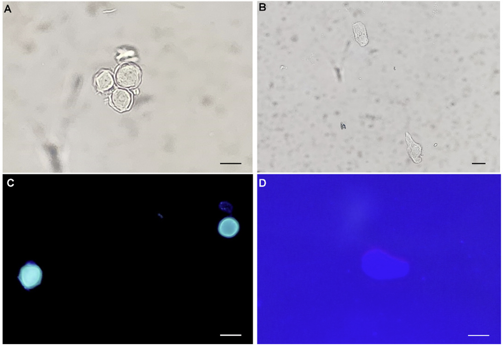
Fig. 2. Morphological analysis of Acanthamoeba spp. using calcofluor-white staining. (A) Acanthamoeba cysts in a group under light microscopy (400×, scale bar = 10 μm). (B) Acanthamoeba trophozoites with a single large nucleus and numerous vacuoles were conspicuous under light microscopy (200×, scale bar = 10 μm). (C) Remarkable Acanthamoeba cysts with double cyst walls under fluorescence microscopy (400×, scale bar = 10 μm). (D) Acanthamoeba trophozoites were relatively unremarkable under fluorescence microscopy (400×, scale bar = 10 μm).
Reference
-
1. Lorenzo-Morales J, Khan NA, Walochnik J. An update on Acanthamoeba keratitis: diagnosis, pathogenesis and treatment. Parasite 2015;22:10. .2. Schroeder JM, Booton GC, Hay J, Niszl IA, Seal DV, Markus MB, et al. Use of subgenic 18S ribosomal DNA PCR and sequencing for genus and genotype identification of Acanthamoebae from humans with keratitis and from sewage sludge. J Clin Microbiol 2001;39:1903–11. .3. Jones BR, McGill JI, Steele AD. Recurrent suppurative kerato-uveitis with loss of eye due to infection by Acanthamoeba castellani. Trans Ophthalmol Soc U K 1975;95:210–3. .4. Lee JE, Hahn TW, Oum BS, Choi HY, Yu HS, Lee JS. Acanthamoeba keratitis related to orthokeratology. Int Ophthalmol 2007;27:45–9. .5. Lee JS, Hahn TW, Choi SH, Yu HS, Lee JE. Acanthamoeba keratitis related to cosmetic contact lenses. Clin Exp Ophthalmol 2007;35:775–7. .6. Kong HH, Shin JY, Yu HS, Kim J, Hahn TW, Hahn YH, et al. Mitochondrial DNA restriction fragment length polymorphism (RFLP) and 18S small-subunit ribosomal DNA PCRRFLP analyses of Acanthamoeba isolated from contact lens storage cases of residents in southwestern Korea. J Clin Microbiol 2002;40:1199–206. .7. Yoon HY and Jeon HS. Diagnosis and management of Acanthamoeba keratitis. Ann Optom Contact Lens 2022;21:104–8. .8. Chan LL, Mak JW, Low YT, Koh TT, Ithoi I, Mohamed SM. Isolation and characterization of Acanthamoeba spp. from air-conditioners in Kuala Lumpur, Malaysia. Acta Trop 2011;117:23–30. .9. Thomas PA and Kuriakose T. Rapid detection of Acanthamoeba cysts in corneal scrapings by lactophenol cotton blue staining. Arch Ophthalmol 1990;108:168. .10. Mackay IM. Real-time PCR in the microbiology laboratory. Clin Microbiol Infect 2004;10:190-212. .11. Qvarnstrom Y, Visvesvara GS, Sriram R, da Silva AJ. Multiplex real-time PCR assay for simultaneous detection of Acanthamoeba spp., Balamuthia mandrillaris, and Naegleria fowleri. J Clin Microbiol 2006;44:3589–95. .
- Full Text Links
-
- Actions
-
Cited
- CITED
-
- Close
- Share
-
- Similar articles
-
- Comparison of Usefulness of Laboratory Diagnosis in Ancanthamoeba Keratitis
- Acanthamoeba Keratitis: Microscopic and Ultrastructural Findings: A case report
- In vivo tandem scanning confocal microscopy in acanthamoeba keratitis
- Two Cases of Corneal Toxicity in Acanthamoeba Keratitis by Combined Topical Anti-Acanthamoeba Keratitis Eye Solution
- Acanthoamoeba Keratitis Induced by a Therapeutic, Soft Contact Lens: Diagnosis via Gram Staining
