J Pathol Transl Med.
2024 Jul;58(4):201-204. 10.4132/jptm.2024.06.07.
What’s new in adrenal gland pathology: WHO 5th edition for adrenal cortex
- Affiliations
-
- 1Department of Laboratory Medicine and Pathology, University of Washington, Seattle, WA, USA
- KMID: 2557761
- DOI: http://doi.org/10.4132/jptm.2024.06.07
Abstract
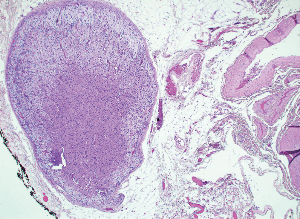
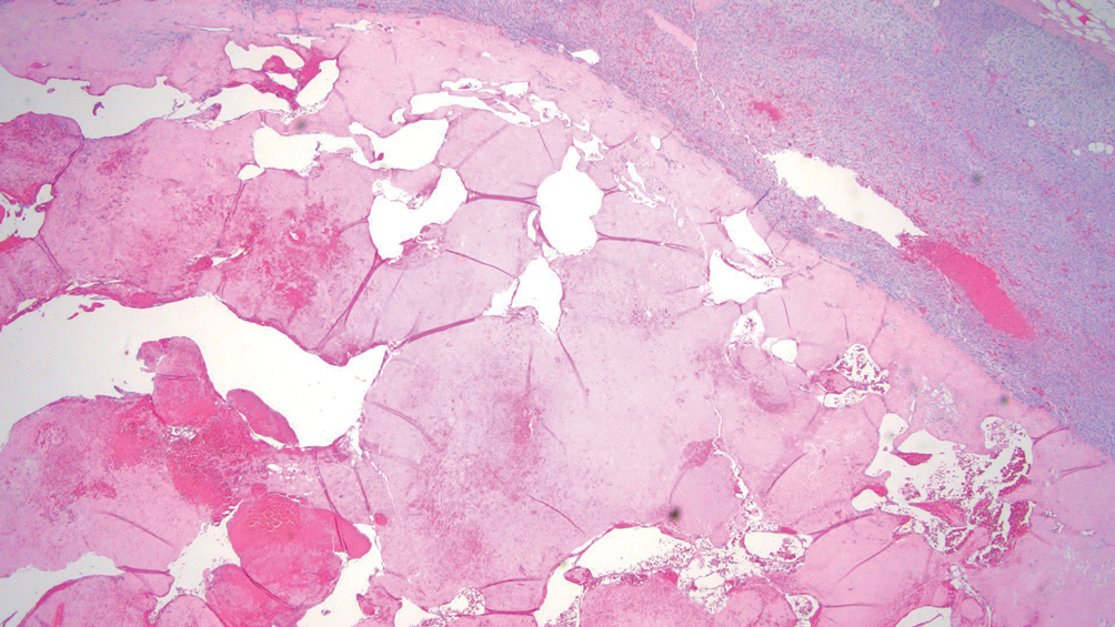
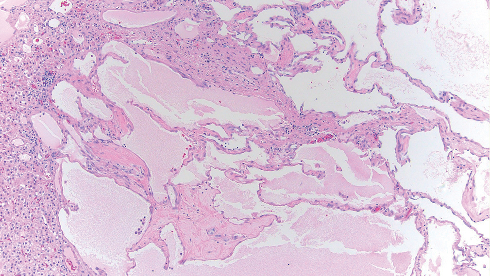
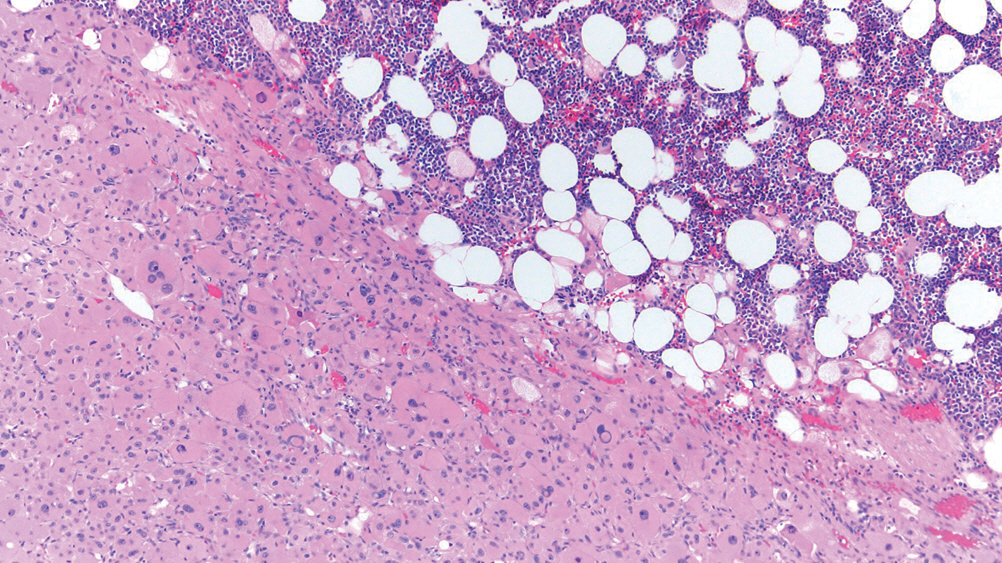
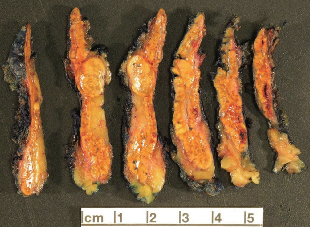
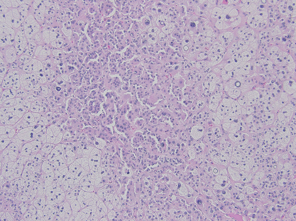
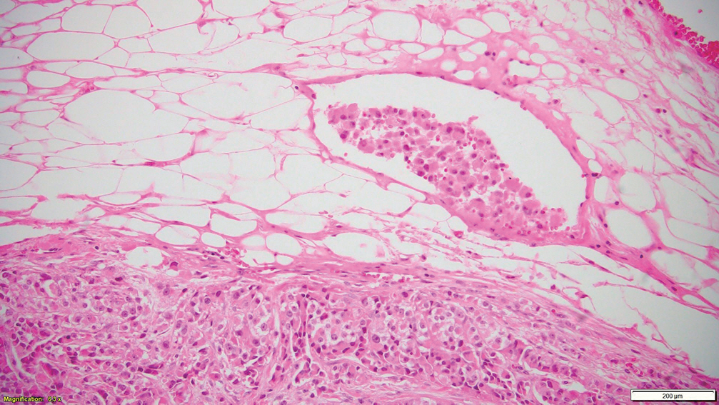
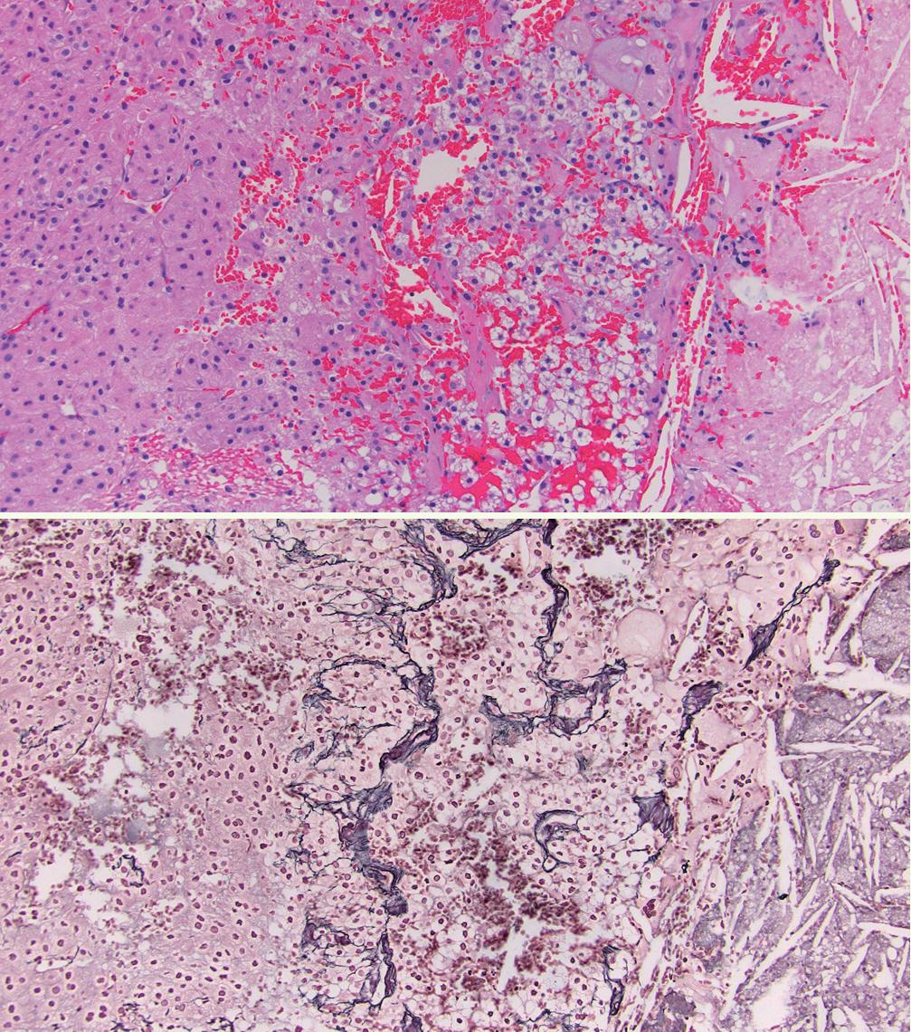
- The 5th edition of WHO Classification of Endocrine and Neuroendocrine Tumors (2022) introduced many significant changes relevant to endocrine daily practice. In this newsletter, we summarize the notable changes to the adrenal cortex based on the 5th edition of the WHO classification [1].
Figure
- Full Text Links
- Actions
-
Cited
- CITED
-
- Close
- Share
- Similar articles
-
- Embryonic Development and Adult Regeneration of the Adrenal Gland
- Radiological Findings of Congenital Lipoid Adrenal Hyperplasia: A Case Report
- A case of primary bilateral adrenal lymphoma with partial adrenal insufficiency
- Nonfunctioning Carcinoma of Adrenal Cortex: A Case Report
- A Case of Congenital Lipoid Adrenal Hyperplasia: Early Diagnosis by Using Computed Tomography

