Heterotopic ovarian hilus cells of the salpinx: a case report and literature review
- Affiliations
-
- 1Department of Pathology, Inje University Haeundae Paik Hospital, Inje University College of Medicine, Busan, Korea
- KMID: 2556800
- DOI: http://doi.org/10.7180/kmj.23.120
Abstract
- Ovarian hilus cells (OHCs), a counterpart of testicular Leydig cells, are usually found in the ovarian poles and produce androstenedione. Their origin remains a matter of debate, although OHCs are assumed to come from the adrenogenital primordium. OHCs are rarely observed around the poles of the ovary, including the mesoovarium, stroma (perisalpinx) of the salpinx, and the wall of paratubal cysts. Their clinical and pathological characteristics are not well-known because of their rarity. Herein, we present a case of ectopic OHCs in a 48-year-old woman. The patient underwent total hysterectomy and bilateral salpingectomy for vaginal bleeding due to multiple leiomyomas. We incidentally found OHCs in the stroma of the infundibulum of the salpinx, just beneath the tubal epithelium. Their size was less than 1 mm, and they were composed of large cells with central round nuclei and abundant clear or granular cytoplasm. OHCs share morphological and immunohistochemical profiles with ectopic adrenal glands, and the differential diagnosis is sometimes difficult. They do not exhibit microscopic encapsulation or the normal adrenal cortex zonation pattern. The patient was discharged and did not show any abnormal findings during 19 months of follow-up. Analyzing the characteristics of testicular Leydig cells will help understand how OHCs develop and why heterotopic OHCs occur in and around the salpinges.
Figure
-
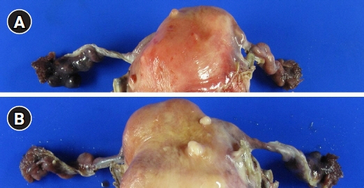
Fig. 1. Gross findings. Anterior (A) and posterior side (B) of the fresh right fallopian tube following hysterectomy with bilateral salpingectomy showing several fimbrial cysts at the end of the right fallopian tube. The external surface of the bilateral salpinges was smooth. A couple of subserosal small leiomyomas were also noted.
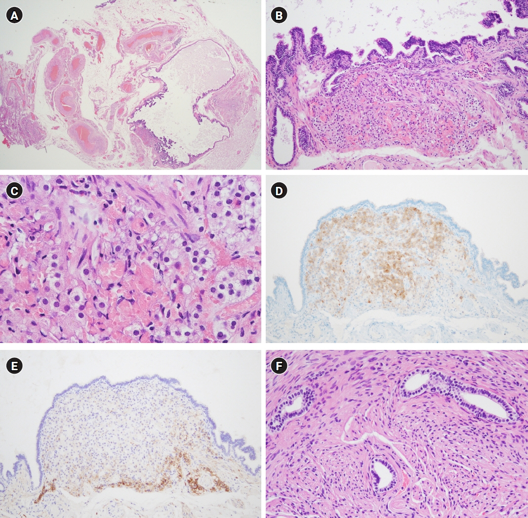
Fig. 2. Microscopic and immunohistochemical findings. (A) Under low-power magnification, the infundibulum of the fallopian tube had a thinned myosalpinx and tertiary branch of plicae. Thick-walled vessels and bundles of nonmyelinated nerves were noted in the mesosalpinx (hematoxylin and eosin stain [H&E], ×12.5). (B) Some nests of epithelial cells are present just beneath the tubal epithelium and admixed with smooth muscle fascicles of the myosalpinx. These oval cells had abundant eosinophilic cytoplasm and were arranged in a trabecular pattern with sinusoidal blood vessels. They were just next to the blood vessels (H&E, ×100). (C) Oval or round cells had round nuclei, some of which had inconspicuous nucleoli. Cells on the left side showed vacuolated cytoplasm (H&E, ×400). (D) The cells were immunoreactive to antibodies produced against inhibin-alpha (inhibin-alpha immunohistochemistry, ×100). (E) CD56 was expressed in the periphery of the nest of hilus cells (CD56 immunohistochemistry, ×100). (F) Wolffian duct remnants, showing ducts lined by cuboidal cells and wrapped with smooth muscle, were identified in the mesosalpinx around ectopic hilus cells (H&E, ×200).
Reference
-
References
1. Vang R. Diseases of the fallopian tube and paratubal region. In : Kurman RJ, Ellenson LH, Ronnet BM, editors. Blaustein's pathology of the female genital tract. Springer;2019. p. 649–714.2. Merrill JA. Ovarian hilus cells. Am J Obstet Gynecol. 1959; 78:1258–71.3. Sternberg WH. The morphology, androgenic function, hyperplasia, and tumors of the human ovarian hilus cells. Am J Pathol. 1949; 25:493–521.4. Lewis JD. Hilus-cell hyperplasia of ovaries and tubes: report of a case. Obstet Gynecol. 1964; 24:728–31.5. Carrasco-Juan JL, Alvarez-Arguelles-Cabrera H, Martin-Corriente C, Gutierrez-Garcia R, Vega-Falcon A, Exposito-Afonso I, et al. Extraparenchymal ovarian and testicular Leydig cells: ectopic/heterotopic or orthotopic? Arch Gynecol Obstet. 2018; 298:655–61.6. Carrasco-Juan JL, Alvarez-Arguelles Cabrera H, Martin Corriente MD, Valladares Parrilla F, Gutierrez Garcia R, Diaz-Flores Feo L. Ovarian Leydig cells (OLC): about their heterotopia or eutopia. Int J Gynecol Pathol. 2018; 37:536.7. Honore LH, O’Hara KE. Ovarian hilus cell heterotopia. Obstet Gynecol. 1979; 53:461–4.8. Carrasco-Juan JL, Alvarez-Arguelles Cabrera H, Martin Corriente MC, Gonzalez-Gomez M, Valladares Parrilla R, Gutierrez Garcia R, et al. Ovarian Leydig cells (OLC): a histomorphological and immunohistochemical study. Histol Histopathol. 2017; 32:1089–97.9. Wen Q, Cheng CY, Liu YX. Development, function and fate of fetal Leydig cells. Semin Cell Dev Biol. 2016; 59:89–98.10. DeFalco T, Takahashi S, Capel B. Two distinct origins for Leydig cell progenitors in the fetal testis. Dev Biol. 2011; 352:14–26.11. Palomaki JF, Blair OM. Hilus cell rest of the fallopian tube: a case report. Obstet Gynecol. 1971; 37:60–2.12. Usubutun A. Hilus cells in tuba uterina; an uncommon localisation and coincidence with endometrial carcinoma: a case report and review of the literature. Turk J Pathol. 2007; 23:52–5.13. Hirschowitz L, Salmons N, Ganesan R. Ovarian hilus cell heterotopia. Int J Gynecol Pathol. 2011; 30:46–52.14. Russell P, Hyne S, Valmadre S. The tubal fimbria: the old broom sweeps into prominence. Pathology. 2013; 45:706–8.15. Ansari IA, Prakash G, K R, V E, Bhat S. Hilus cell hyperplasia of fallopian tubes: a rare and incidental finding with uterine leiomyomas. Kathmandu Univ Med J (KUMJ). 2014; 12:64–6.16. He HL, Lee YE, Chang CC. Hilus cell heterotopia accompanying bilateral ovarian serous cystadenomas: a case report and review of the literature. Int J Clin Exp Pathol. 2014; 7:1246–9.17. Chougule A, Dey P. Hilus cell heterotopia with intraneural eutopia. Int J Gynecol Pathol. 2017; 36:200–2.18. Camacho-Partida IG, Ortiz-Hidalgo C. Hilus cell heterotopia mimicking perineurial invasion in a woman with endometrial adenocarcinoma. Patología. 2019; 57:1–6.19. Hu YH, Yu CT, Wen MC. Hilus cell heterotopia of fallopian tube: a rare and incidental finding with high grade squamous intraepithelial lesion of cervix. J Obstet Gynaecol. 2020; 40:1031–3.20. Hatano O, Takakusu A, Nomura M, Morohashi K. Identical origin of adrenal cortex and gonad revealed by expression profiles of Ad4BP/SF-1. Genes Cells. 1996; 1:663–71.
- Full Text Links
-
- Actions
-
Cited
- CITED
-
- Close
- Share
-
- Similar articles
-
- Ovarian Rete Cyst in a Post-menopausal Woman: A Case Report
- Calcinosis circumscripta of the both salpinx: A case report
- A case of heterotopic pregnancy after ovulation induction and intrauterine insemination
- A Case of Discovery of Heterotopic Pregnancy After Elective Abortion
- A Case of Heterotopic Pregnancy in a Natural Cycle
