J Neurocrit Care.
2024 Jun;17(1):34-35. 10.18700/jnc.240002.
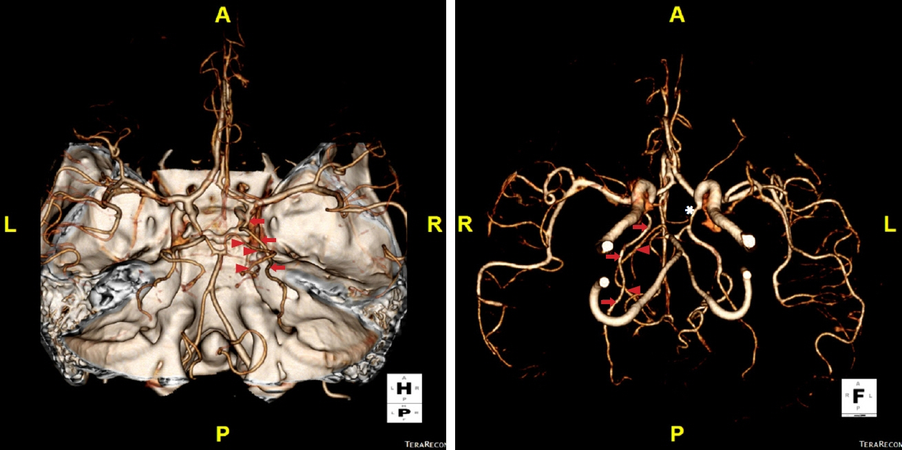
Accessory posterior cerebral artery (PCA): a rare variant of PCA
- Affiliations
-
- 1Department of Neurology, Sanggye Paik Hospital, Inje University College of Medicine, Seoul, Korea
- 2Department of Neurology, Bundang Jesaeng General Hospital, Seongnam, Korea
- KMID: 2556732
- DOI: http://doi.org/10.18700/jnc.240002
Figure
Reference
-
1. Uchino A, Saito N, Takahashi M, Okano N, Tanisaka M. Variations of the posterior cerebral artery diagnosed by MR angiography at 3 tesla. Neuroradiology. 2016; 58:141–6.2. Takahashi S, Suga T, Kawata Y, Sakamoto K. Anterior choroidal artery: angiographic analysis of variations and anomalies. AJNR Am J Neuroradiol. 1990; 11:719–29.3. Rusu MC, Vrapciu AD, Lazăr M. A rare variant of accessory posterior cerebral artery. Surg Radiol Anat. 2023; 45:523–6.
- Full Text Links
- Actions
-
Cited
- CITED
-
- Close
- Share
- Similar articles
-
- Microsurgical Anatomy of the Basilar Artery and Posterior Cerebral Artery
- A Case of Bilateral Posterior Cerebral Artery Infarction with Patent Foramen Ovale
- Giant Serpentine Aneurysm of the Posterior Cerebral Artery: Case Report
- The Management and Characteristics of Posterior Cerebral Artery Aneurysms
- Novel Superior Cerebellar Artery Aneurysm Coming from a Superior Cerebellar Artery-Posterior Cerebral Artery Anastomotic Branch


