Neurointervention.
2024 Jul;19(2):129-134. 10.5469/neuroint.2024.00136.
Endovascular Management of a Ruptured Aneurysm on a Posterior Inferior Cerebellar Artery with Extradural C2-Origin: Case Report and Literature Review
- Affiliations
-
- 1Department of Radiology, Copenhagen University Hospital - Rigshospitalet, Copenhagen, Denmark
- 2Department of Radiology, Baylor College of Medicine, Houston, TX, USA
- 3Department of Interventional Radiology, The University of Texas MD Anderson Cancer Center, Houston, TX, USA
- 4Department of Neurosurgery, Baylor College of Medicine, Houston, TX, USA
- KMID: 2556666
- DOI: http://doi.org/10.5469/neuroint.2024.00136
Abstract
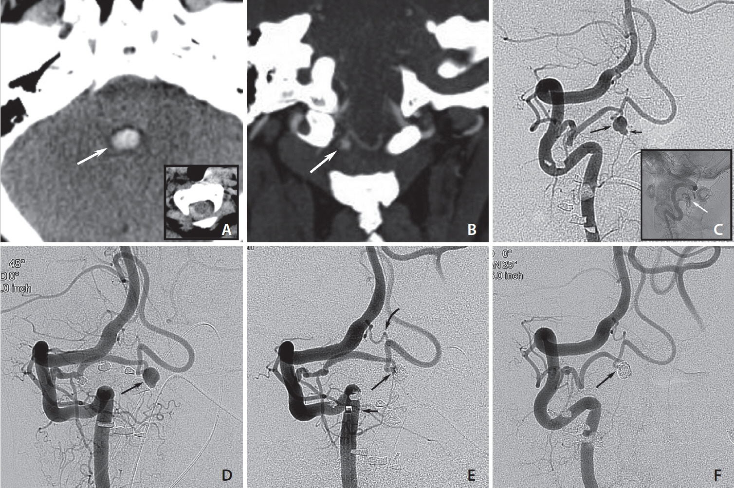
- Extracranial vascular pathology uncommonly causes intracranial subarachnoid hemorrhage (SAH). Among possible lesions are aneurysms at the craniocervical junction arising from a posterior inferior cerebellar artery (PICA) with an extradural origin. We describe a case of a 55-year-old female presenting with a sudden and severe headache. A computed tomography scan revealed a SAH within the fourth ventricle and cervical spinal canal, and a ruptured saccular aneurysm on a PICA with extradural C2-origin. Despite difficult access anatomy, endovascular treatment was feasible and resulted in subtotal initial occlusion and preservation of distal PICA flow. Upon 3-month follow-up, the aneurysm was completely occluded with a patent PICA. The patient’s clinical status remained stable at the 1.5-year follow-up. In conclusion, we present a rare case of an aneurysm originating from a PICA with extradural C2-origin that was treated endovascularly with preservation of the PICA.
Figure
Reference
-
1. Dammers R, Krisht AF, Partington S. Diagnosis and surgical management of extracranial PICA aneurysms presenting through subarachnoid haemorrhage: case report and review of the literature. Clin Neurol Neurosurg. 2009; 111:758–761.2. Fine AD, Cardoso A, Rhoton AL Jr. Microsurgical anatomy of the extracranial-extradural origin of the posterior inferior cerebellar artery. J Neurosurg. 1999; 91:645–652.3. Abe T, Kojima K, Singer RJ, Marks MP, Watanabe M, Ohtsuru K, et al. Endovascular management of an aneurysm arising from posterior inferior cerebellar artery originated at the level of C2. Radiat Med. 1998; 16:141–143.4. Tabatabai SA, Zadeh MZ, Meybodi AT, Hashemi M. Extracranial aneurysm of the posterior inferior cerebellar artery with an aberrant origination: case report. Neurosurgery. 2007; 61:E1097–1098. discussion E1098.5. Lasjaunias P, Vallee B, Person H, Ter Brugge K, Chiu M. The lateral spinal artery of the upper cervical spinal cord. Anatomy, normal variations, and angiographic aspects. J Neurosurg. 1985; 63:235–241.6. Siclari F, Burger IM, Fasel JH, Gailloud P. Developmental anatomy of the distal vertebral artery in relationship to variants of the posterior and lateral spinal arterial systems. AJNR Am J Neuroradiol. 2007; 28:1185–1190.7. Dikshit P, Sharma A, Mehrotra A, Verma PK, Das KK, Jaiswal AK. Extra-cranial proximal pica aneurysm - a rare and surreptious cause of posterior fossa sah: case report and review of literature. [published online ahead of print Sep 15, 2021]. Br J Neurosurg. 2021.8. Bhat DI, Somanna S, Kovoor J, Chandramoul BA. Aneurysms from extracranial, extradurally originating posterior inferior cerebellar arteries: a rare case report. Surg Neurol. 2009; 72:406–408.
Article9. Tanaka A, Kimura M, Yoshinaga S, Tomonaga M. Extracranial aneurysm of the posterior inferior cerebellar artery: case report. Neurosurgery. 1993; 33:742–744. discussion 744-745.
Article10. Kim K, Kobayashi S, Mizunari T, Teramoto A. Aneurysm of the distal posteroinferior cerebellar artery of extracranial origin: case report. Neurosurgery. 2001; 49:996–998. discussion 998-999.
Article11. Kleinpeter G. Why are aneurysms of the posterior inferior cerebellar artery so unique? Clinical experience and review of the literature. Minim Invasive Neurosurg. 2004; 47:93–101.
Article12. Lister JR, Rhoton AL Jr, Matsushima T, Peace DA. Microsurgical anatomy of the posterior inferior cerebellar artery. Neurosurgery. 1982; 10:170–199.13. Margolis MT, Newton TH. Borderlands of the normal and abnormal posterior inferior cerebellar artery. Acta Radiol Diagn (Stockh). 1972; 13:163–176.
Article14. Wakao N, Takeuchi M, Nishimura M, Riew KD, Kamiya M, Hirasawa A, et al. Vertebral artery variations and osseous anomaly at the C1-2 level diagnosed by 3D CT angiography in normal subjects. Neuroradiology. 2014; 56:843–849.
Article15. Salas E, Ziyal IM, Bank WO, Santi MR, Sekhar LN. Extradural origin of the posteroinferior cerebellar artery: an anatomic study with histological and radiographic correlation. Neurosurgery. 1998; 42:1326–1331.
Article16. Lasjaunias P, Berenstein A, ter Brugge KG. Craniocervical junction. In: Lasjaunias P, Berenstein A, Brugge KG. Surgical neuroangiography, 2nd ed. Vol. 1, Clinical vascular anatomy and variations. Springer, 2001;165-259.17. Won D, Lee JM, Park IS, Lee CH, Lee K, Kim JY, et al. Posterior inferior cerebellar artery infarction originating at C1-2 after C1-2 fusion. Korean J Neurotrauma. 2019; 15:192–198.
Article
- Full Text Links
- Actions
-
Cited
- CITED
-
- Close
- Share
- Similar articles
-
- Successful Endovascular Treatment of Ruptured Superior Cerebellar Artery Aneurysm Associated with Moyamoya Disease : A Case Report and Review of the Literature
- Stent-Jack Technique for Ruptured Vertebral Artery Dissecting Aneurysm Involving the Origin of Posterior Inferior Cerebellar Artery
- Aneurysm of the Posteior Inferior Cerebellar Artery Associated with Arteriovenous Malformation: Case Report
- Aneurysm of Distal Posterior Inferior Cerebellar Artery:Case Report with Review of the Literature
- Dissecting Aneurysm Associated with a Double Origin of the Posterior Inferior Cerebellar Artery Causing Subarachnoid Hemorrhage


