Neurointervention.
2024 Jul;19(2):111-117. 10.5469/neuroint.2024.00157.
Treatment of Traumatic Direct Carotid-Cavernous Fistula with a BeGraft-Covered Stent
- Affiliations
-
- 1Department of Neurosurgical, Section of Neurovascular, Ghaem Hospital, Mashhad University of Medical Sciences, Mashhad, Iran
- 2Department of Neurosurgery, Emam Hospital, Mazandaran University of Medical Sciences, School of Medicine, Sari, Iran
- 3Department of Interventional Neuroradiology, Rothschild Foundation Hospital, Paris, France
- 4Department of Neurosurgery, Firouzgar Hospital, Iran University of Medical Sciences, School of Medicine, Tehran, Iran
- 5Division of Stroke and Endovascular Neurosurgery, Department of Neurological Surgery, Keck School of Medicine, University of Southern California (USC), Los Angeles, CA, USA
- KMID: 2556663
- DOI: http://doi.org/10.5469/neuroint.2024.00157
Abstract
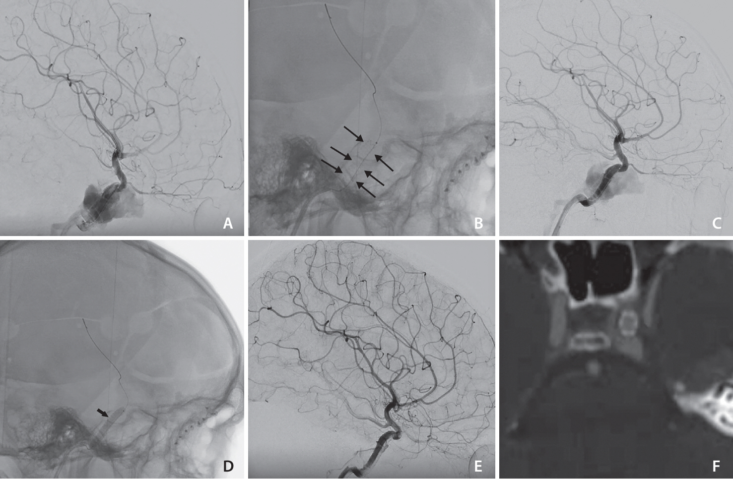
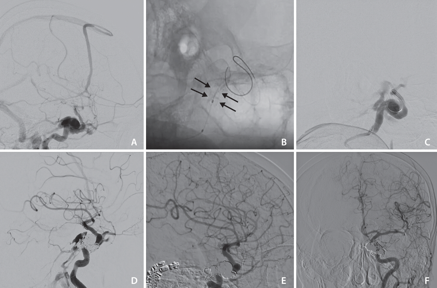
- The widely accepted option for treating traumatic direct carotid-cavernous fistula (dCCF) has been endovascular treatment using detachable balloons, coils, or embolic agents. Covered stent deployment has been applied by a few operators and has shown promising results. This is a retrospective study on patients with dCCF treated by an endovascular approach using BeGraft, a covered stent. In 4 cases, this device was successfully deployed without any complications. Immediate complete occlusion was achieved in 3 patients (75%) after deployment of the covered stents. One patient required transvenous coiling for occlusion of the remaining endoleak. Follow-up imaging demonstrated 100% fistula occlusion with complete internal carotid artery patency. No early or late complications occurred following treatment. In conclusion, the BeGraft-covered stent could be a promising safe and effective alternative option for the endovascular treatment of dCCF.
Figure
Reference
-
1. Serbinenko FA. Balloon catheterization and occlusion of major cerebral vessels. J Neurosurg. 1974; 41:125–145.
Article2. Ide S, Kiyosue H, Tokuyama K, Hori Y, Sagara Y, Kubo T. Direct carotid cavernous fistulas. J Neuroendovasc Ther. 2020; 14:583–592.
Article3. Texakalidis P, Tzoumas A, Xenos D, Rivet DJ, Reavey-Cantwell J. Carotid cavernous fistula (CCF) treatment approaches: a systematic literature review and meta-analysis of transarterial and transvenous embolization for direct and indirect CCFs. Clin Neurol Neurosurg. 2021; 204:106601.
Article4. Farhatnia Y, Tan A, Motiwala A, Cousins BG, Seifalian AM. Evolution of covered stents in the contemporary era: clinical application, materials and manufacturing strategies using nanotechnology. Biotechnol Adv. 2013; 31:524–542.
Article5. Lu D, Ma T, Zhu G, Zhang T, Wang N, Lei H, et al. Willis covered stent for treating intracranial pseudoaneurysms of the internal carotid artery: a multi-institutional study. Neuropsychiatr Dis Treat. 2022; 18:125–135.
Article6. Yin B, Sheng HS, Wei RL, Lin J, Zhou H, Zhang N. Comparison of covered stents with detachable balloons for treatment of posttraumatic carotid-cavernous fistulas. J Clin Neurosci. 2013; 20:367–372.
Article7. Liu Q, Qi C, Wang Y, Su W, Li G, Wang D. Treatment of direct carotid-cavernous fistula with Willis covered stent with midterm follow-up. Chin Neurosurg J. 2021; 7:41.
Article8. Li J, Lan ZG, Xie XD, You C, He M. Traumatic carotid-cavernous fistulas treated with covered stents: experience of 12 cases. World Neurosurg. 2010; 73:514–519.
Article9. Archondakis E, Pero G, Valvassori L, Boccardi E, Scialfa G. Angiographic follow-up of traumatic carotid cavernous fistulas treated with endovascular stent graft placement. AJNR Am J Neuroradiol. 2007; 28:342–347.10. Gomez F, Escobar W, Gomez AM, Gomez JF, Anaya CA. Treatment of carotid cavernous fistulas using covered stents: midterm results in seven patients. AJNR Am J Neuroradiol. 2007; 28:1762–1768.
Article11. Li K, Cho YD, Kim KM, Kang HS, Kim JE, Han MH. Covered stents for the endovascular treatment of a direct carotid cavernous fistula: single center experiences with 10 cases. J Korean Neurosurg Soc. 2015; 57:12–18.
Article12. Wang W, Li MH, Li YD, Gu BX, Lu HT. Reconstruction of the internal carotid artery after treatment of complex traumatic direct carotid-cavernous fistulas with the willis covered stent: a retrospective study with long-term follow-up. Neurosurgery. 2016; 79:794–805.
Article13. Wang C, Xie X, You C, Zhang C, Cheng M, He M, et al. Placement of covered stents for the treatment of direct carotid cavernous fistulas. AJNR Am J Neuroradiol. 2009; 30:1342–1346.
Article14. Wang W, Li YD, Li MH, Tan HQ, Gu BX, Wang J, et al. Endovascular treatment of post-traumatic direct carotid-cavernous fistulas: a single-center experience. J Clin Neurosci. 2011; 18:24–28.
Article15. He XH, Li WT, Peng WJ, Lu JP, Liu Q, Zhao R. Endovascular treatment of posttraumatic carotid-cavernous fistulas and pseudoaneurysms with covered stents. J Neuroimaging. 2014; 24:287–291.
Article16. Petrov I, Stankov Z, Boychev D, Klissurski M. Use of coronary stent grafts for the treatment of high-flow carotid cavernous fistula. BMJ Case Rep. 2021; 14:e245922.
Article17. Jeong SH, Lee JH, Choi HJ, Kim BC, Yu SH, Lee JI. First line treatment of traumatic carotid cavernous fistulas using covered stents at level 1 regional trauma center. J Korean Neurosurg Soc. 2021; 64:818–826.
Article18. Wroe WW, Zeineddine HA, Dawes BH, Martinez-Gutierrez JC, Shah M, Spiegel G, et al. Treatment of traumatic direct carotid cavernous fistula with a PK papyrus covered stent: a report of 2 cases. Stroke Vasc Interv Neurol. 2023; 3:e001015.
Article19. Kilic ID, Fabris E, Serdoz R, Caiazzo G, Foin N, Abou-Sherif S, et al. Coronary covered stents. EuroIntervention. 2016; 12:1288–1295.
Article20. Kufner S, Schacher N, Ferenc M, Schlundt C, Hoppmann P, Abdel-Wahab M, et al. Outcome after new generation single-layer polytetrafluoroethylene-covered stent implantation for the treatment of coronary artery perforation. Catheter Cardiovasc Interv. 2019; 93:912–920.
Article
- Full Text Links
- Actions
-
Cited
- CITED
-
- Close
- Share
- Similar articles
-
- Regional Cerebral Blood Flow Changes in Traumatic Carotid Cavernous Fistula During Trapping Procedure: Case Study, Preliminary Report
- Traumatic Carotid-cavernous Fistula Bringing about Intracerebral Hemorrhage
- Direct Microsurgical Repair of Traumatic Carotid-Cavernous Fistula
- Treatment of High-Flow Carotid Cavernous Fistula Using a Graft Stent: Case Report
- Bilateral Traumatic Carotid-Cavernous Fistula


