Neurointervention.
2024 Jul;19(2):102-105. 10.5469/neuroint.2024.00094.
A Novel Training Method for Endovascular Clot Retrieval Using a Portable Vascular Model and Red Film
- Affiliations
-
- 1Stroke Center, Aichi Medical University, Aichi, Japan
- 2Neuroendovascular Therapy Center, Aichi Medical University, Aichi, Japan
- 3Department of Neurological Surgery, Aichi Medical University, Aichi, Japan
- KMID: 2556661
- DOI: http://doi.org/10.5469/neuroint.2024.00094
Abstract
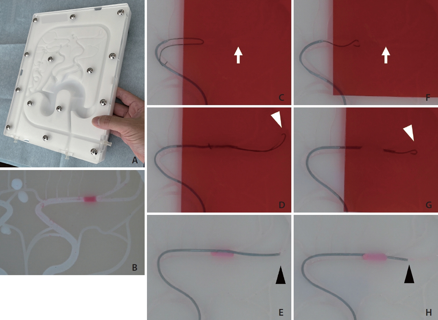
- Hands-on training is a crucial part of education in neuroendovascular treatment to ensure safe and rapid acquisition of techniques. However, there is a significant gap between training and actual clinical practice. This study will introduce innovations for more practical thrombus retrieval training that was developed in this process. A Smart Vascular Model 3 in 1 was used. A pink pseudothrombus was inserted into the M1 (horizontal segment of the middle cerebral artery) section of the model. Then, a “red underlay” purchased at a stationery store was placed to cover the proximal part of M1 and beyond so that the pseudothrombus was not visible. The thrombus was retrieved during training by looking for the location of the thrombus based on the behavior and resistance of the tip of the guidewire and deployment of the stent retriever. The participants were required to have detailed observation skills and precise manipulation skills using a red film to prevent the direct visualization of the pseudothrombus. The implementation of this innovation to the previous hands-on training made the training more practical and effective. If the exact thrombus location can be determined by the behavior of the wire tip, the device’s capabilities can be maximized, and rapid retrieval can be expected. It could also reduce complications, as unnecessary peripheral guidance of the device could be avoided.
Figure
Reference
-
1. Ohshima T, Koiwai M, Matsuo N, Miyachi S. A novel remote hands-on training for neuroendovascular-based treatment in the era of the COVID-19 pandemic. Interv Neuroradiol. 2023; 29:43–46.
Article2. Ato F, Ohshima T, Miyachi S, Matsuo N, Kawaguchi R, Takayasu M. Efficacy and safety of a modified pigtail-shaped microguidewire during endovascular thrombectomy. Asian J Neurosurg. 2019; 14:1040–1043.3. Ohshima T, Kawaguchi R, Maejima R, Yukiue N, Matsuo N, Miyachi S. Initial clinical experience of using a newly designed preshaped microguidewire in acute endovascular thrombectomy. Asian J Neurosurg. 2020; 15:241–244.
Article
- Full Text Links
- Actions
-
Cited
- CITED
-
- Close
- Share
- Similar articles
-
- Feasibility Study of Utilizing a Portable Spectrophotometer to Assess the Radiation Dose Delivered to the Radiochromic Film
- Surgical Removal of the Inferior Vena Cava Filter Using Minimal Cavotomy: A Case Report
- Local Complications Associated with Vascular and Endovascular Surgery
- Spaced Retrieval Effects in Older Adults with Mild Alzheimer's Disease
- Evolution of Endovascular Therapy in Acute Stroke: Implications of Device Development


