Relationship to the superficial radial nerve and anatomic variations of the first extensor compartment in Thai population: a basis for successful de Quervain tenosynovitis treatment
- Affiliations
-
- 1Doctor of Medicine Program, Faculty of Medicine, Chulalongkorn University, Bangkok, Thailand
- 2Department of Anatomy, Faculty of Medicine, Chulalongkorn University, Bangkok, Thailand
- KMID: 2556569
- DOI: http://doi.org/10.5115/acb.24.011
Abstract
- Knowledge of the superficial radial nerve (SRN) relationship and anatomic variations of the first extensor compartment (1st EC) will contribute to a better outcome of de Quervain tenosynovitis treatment. We dissected 87 embalmed cadaveric wrists to determine the relationship of the SRN, the 1st EC length, distance from the proximal and distal 1st EC borders to radial styloid process (RSP), abductor pollicis longus (APL) and extensor pollicis brevis (EPB) tendon slip numbers, and the presence of septum. Our results revealed SRN crossing over the 1st EC in 59.5%. The lateral branch of the superficial radial nerve to the 1st EC midline in most cases (61.9%) except for one specimen, where lateral antebrachial cutaneous nerve was the closest. Distances from proximal and distal 1st EC borders to the RSP were 19.7±4.1 mm and 7.6±1.8 mm, respectively. Extensor retinaculum (ER) width over 1st EC (1st EC length) was 14.8±3.2 mm. Complete and incomplete septa were found in 17.2%, and 42.5%, respectively. The most frequent APL tendon slip number in the compartment was two in overall 47 specimens (54.0%). Almost all compartments (85 specimens; 97.7%) contained one EPB tendon slip. We detected bilateral EPB absence in one cadaver. Moreover, we recorded a tendon slip from extensor pollicis longus traveling into 1st EC bilaterally in one cadaver and observed the EPB muscle belly extension into 1st EC in 9 wrists. Awareness of 1st EC anatomic variations would be essential for successful surgical and nonsurgical outcomes.
Figure
-
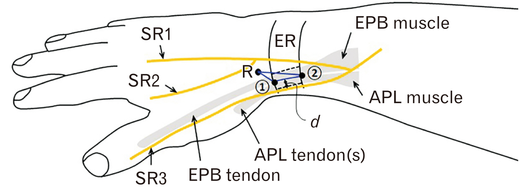
Fig. 1 The diagram of the right wrist demonstrates landmarks and structures investigated in this study. 1, distal border of ER; 2, proximal border of ER; d, distance to the closest branch of superficial radial nerve; APL, abductor pollicis longus; EPB, extensor pollicis brevis; ER, extensor retinaculum; R, radial styloid process; SR, branches of the superficial radial nerve.
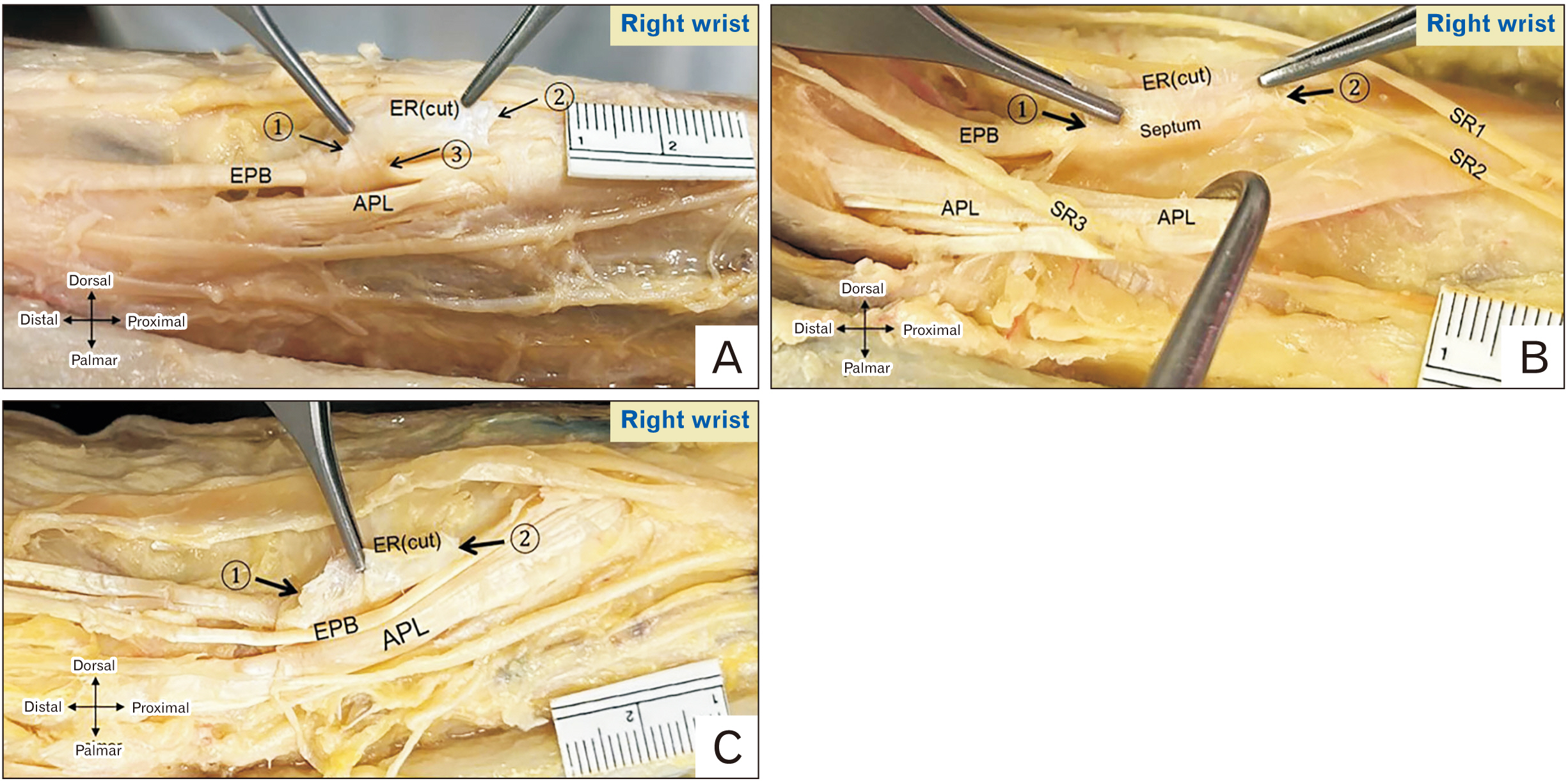
Fig. 2 Types of the first extensor compartment septa. (A) An incomplete septum separating EPB and APL tendons. (B) A complete septum. (C) No septum. 1, distal border of ER; 2, proximal border of ER; 3, proximal border of the incomplete septum; APL, abductor pollicis longus; EPB, extensor pollicis brevis; ER, extensor retinaculum; SR, branches of superficial radial nerve.
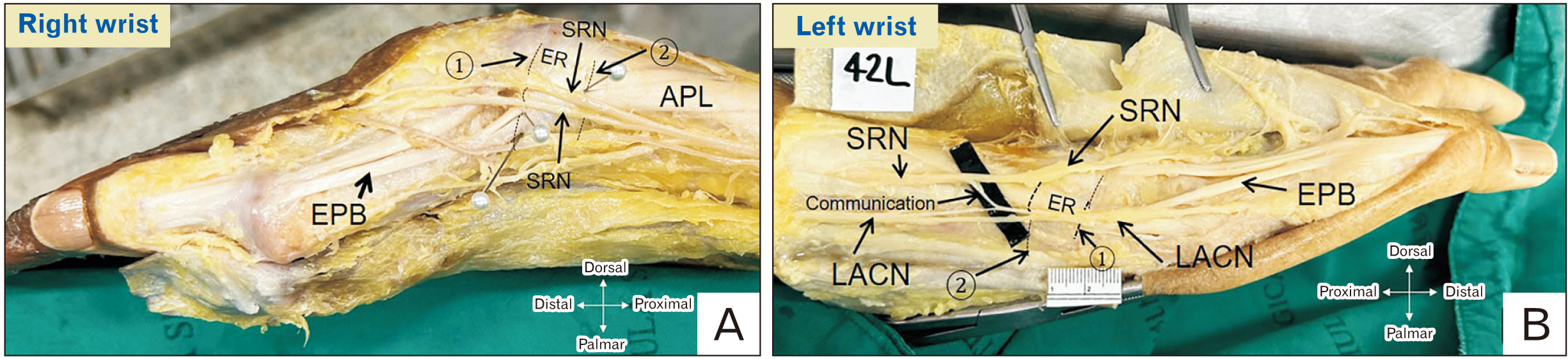
Fig. 3 Relation of the SRN and first EC. (A) Right cadaveric wrist showing two branches of SRN lying on the ER over the first EC. (B) Left cadaveric wrist showing the LACN lying on the ER over the first EC and a communicating branch between SRN and LACN. 1, distal border of ER; 2, proximal border of ER; APL, abductor pollicis longus; ER, extensor retinaculum; EPB, extensor pollicis brevis; LACN, lateral antebrachial cutaneous nerve; SRN, superficial radial nerve; EC, extensor compartment.
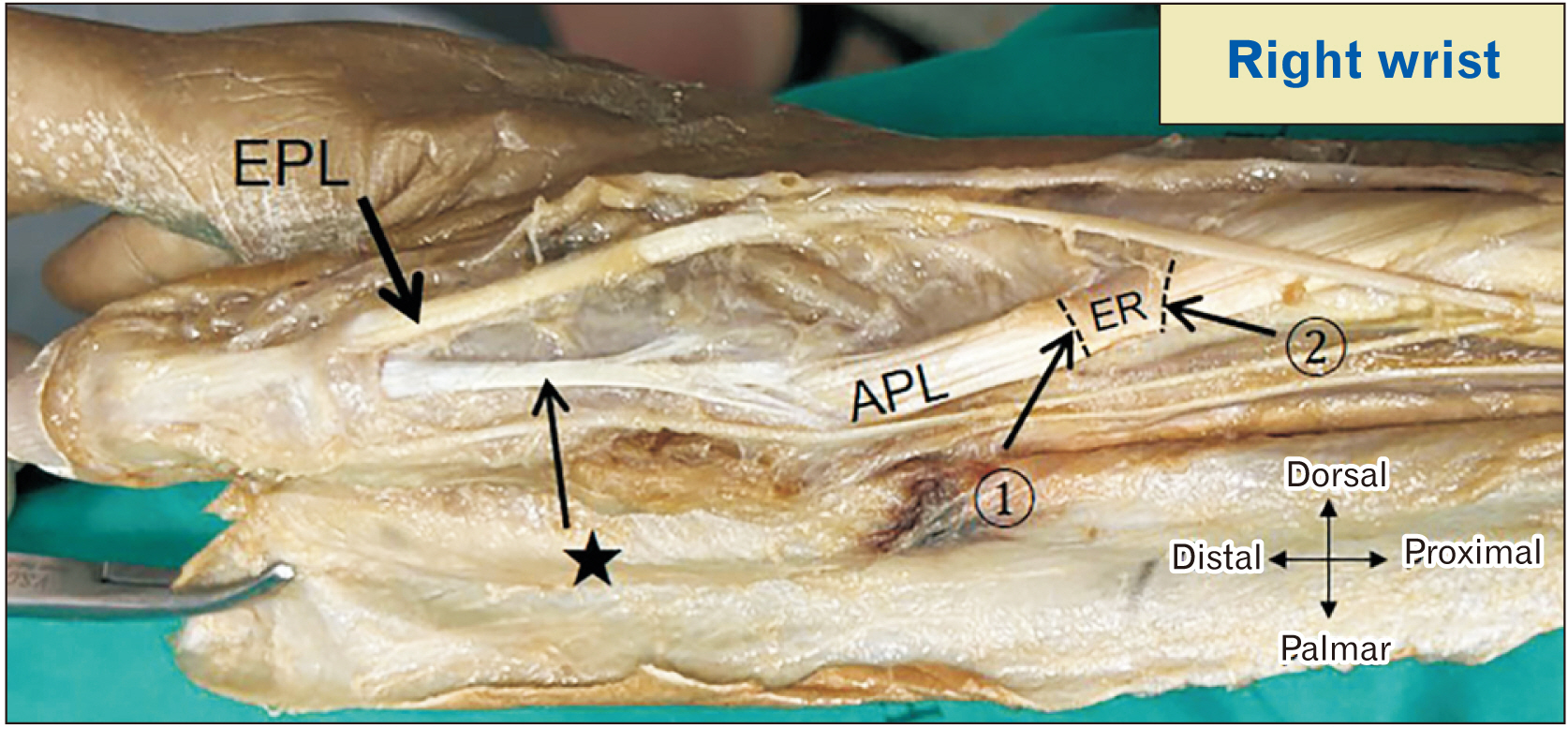
Fig. 4 Absence of EPB in the first extensor compartment. A slip from APL (asterisk) inserting at the proximal phalanx. EPB, extensor pollicis brevis; APL, abductor pollicis longus; EPL, extensor pollicis longus; ER, extensor retinaculum; 1, distal border of ER; 2, proximal border of ER.
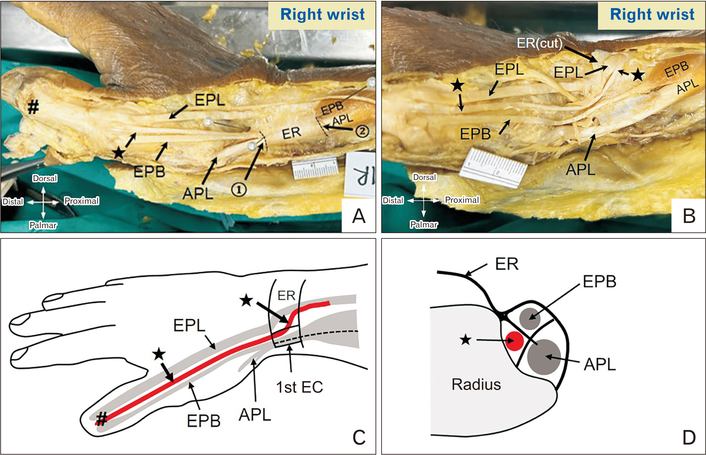
Fig. 5 Photographs and diagrams illustrating an EPL split tendon and three sub-compartments within the first EC. (A) Before ER cut, two tendons distal to the first EC. (B) After the ER cut, one EPB tendon and a split tendon from EPL (asterisk). (C) Course of the split EPL tendon. (D) Cross-section of the 1st EC showing three sub-compartments and contents. 1, distal border of ER; 2, proximal border of ER; asterisk, split EPL tendon; #common insertion of EPB and EPL at distal phalanx of thumb. APL, abductor pollicis longus tendon; EPB, extensor pollicis brevis tendon; EPL, extensor pollicis longus tendon; ER, extensor retinaculum; EC, extensor compartment.
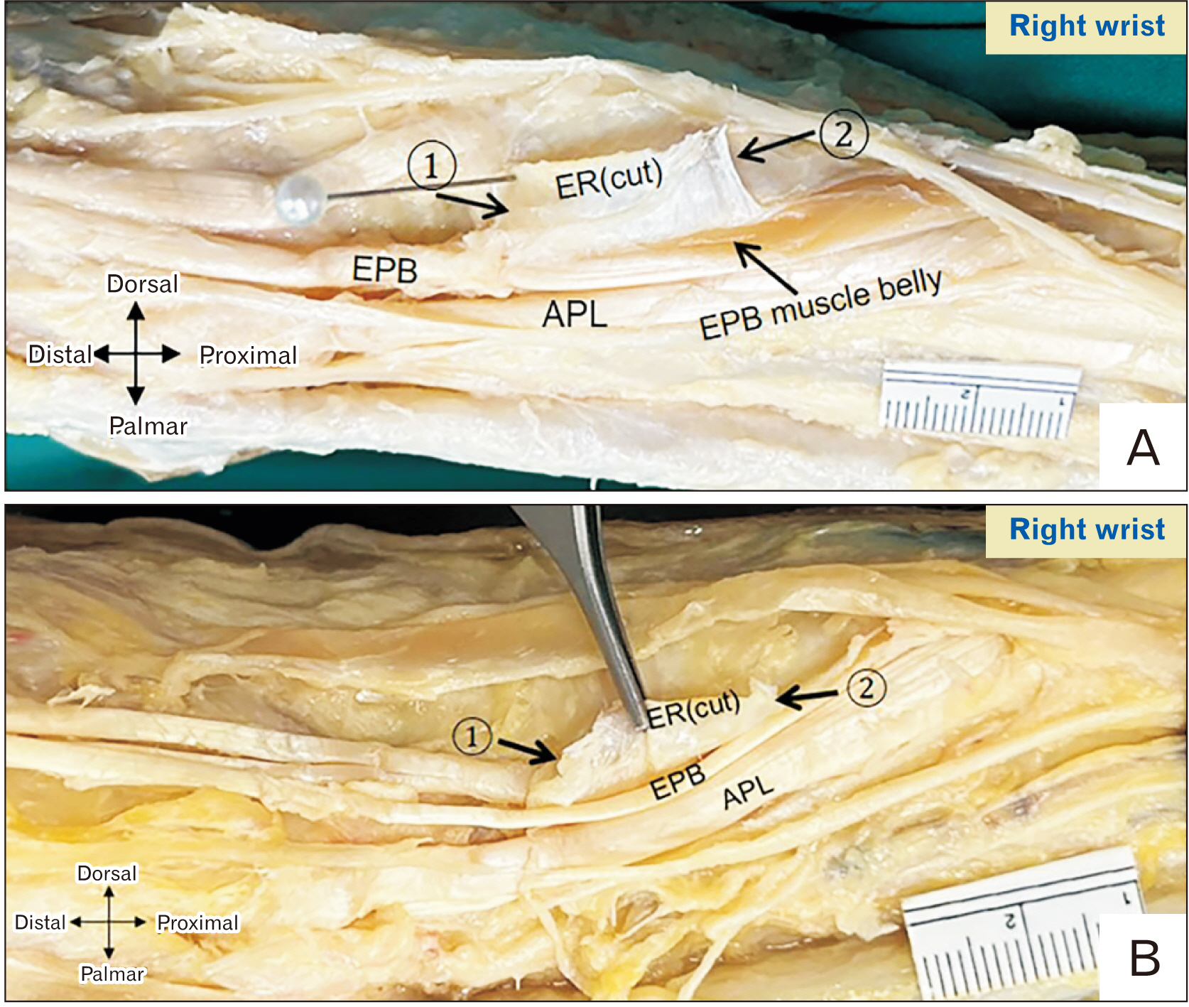
Fig. 6 Illustration of the extension of EPB muscle belly into the first EC. (A) Right cadaveric wrist with the extension of the EPB muscle belly into the first EC. (B) Right cadaveric wrist without extension of EPB muscle belly. 1, distal border of the 1st EC; 2, proximal border of the 1st EC; APL, abductor pollicis longus; EPB, extensor pollicis brevis; EPL, extensor pollicis longus; ER, extensor retinaculum; EC, extensor compartment.
Reference
-
References
1. Lee ZH, Stranix JT, Anzai L, Sharma S. 2017; Surgical anatomy of the first extensor compartment: a systematic review and comparison of normal cadavers vs. De Quervain syndrome patients. J Plast Reconstr Aesthet Surg. 70:127–31. DOI: 10.1016/j.bjps.2016.08.020. PMID: 27693273.
Article2. Currie KB, Tadisina KK, Mackinnon SE. 2022; Common hand conditions: a review. JAMA. 327:2434–45. Erratum in: JAMA 2023;330:772. DOI: 10.1001/jama.2022.8481. PMID: 35762992.3. Ilyas AM, Ast M, Schaffer AA, Thoder J. 2007; De quervain tenosynovitis of the wrist. J Am Acad Orthop Surg. 15:757–64. Erratum in: J Am Acad Orthop Surg 2008;16:35A. DOI: 10.5435/00124635-200712000-00009. PMID: 18063716.
Article4. Larsen CG, Fitzgerald MJ, Nellans KW, Lane LB. 2021; Management of de Quervain tenosynovitis: a critical analysis review. JBJS Rev. 9:e21.00069. DOI: 10.2106/JBJS.RVW.21.00069. PMID: 34506345.5. Harvey FJ, Harvey PM, Horsley MW. 1990; De Quervain's disease: surgical or nonsurgical treatment. J Hand Surg Am. 15:83–7. DOI: 10.1016/S0363-5023(09)91110-8. PMID: 2299173.
Article6. Gurses IA, Coskun O, Gayretli O, Kale A, Ozturk A. 2015; The anatomy of the fibrous and osseous components of the first extensor compartment of the wrist: a cadaveric study. Surg Radiol Anat. 37:773–7. DOI: 10.1007/s00276-015-1439-2. PMID: 25645546.
Article7. Ilyas AM. 2009; Nonsurgical treatment for de Quervain's tenosynovitis. J Hand Surg Am. 34:928–9. DOI: 10.1016/j.jhsa.2008.12.030. PMID: 19410999.
Article8. McDermott JD, Ilyas AM, Nazarian LN, Leinberry CF. 2012; Ultrasound-guided injections for de Quervain's tenosynovitis. Clin Orthop Relat Res. 470:1925–31. DOI: 10.1007/s11999-012-2369-5. PMID: 22552767. PMCID: PMC3369070.
Article9. Güleç A, Türkmen F, Toker S, Acar MA. 2016; Percutaneous release of the first dorsal extensor compartment: a cadaver study. Plast Reconstr Surg Glob Open. 4:e1022. DOI: 10.1097/GOX.0000000000001022. PMID: 27826460. PMCID: PMC5096515.
Article10. Saaiq M. 2021; Management outcome of de Quervain's disease with corticosteroid injection versus surgical decompression. Arch Bone Jt Surg. 9:167–73. DOI: 10.22038/abjs.2020.47822.2359. PMID: 34026933. PMCID: PMC8121040.11. Zingas C, Failla JM, Van Holsbeeck M. 1998; Injection accuracy and clinical relief of de Quervain's tendinitis. J Hand Surg Am. 23:89–96. DOI: 10.1016/S0363-5023(98)80095-6. PMID: 9523961.
Article12. Weller WJ. Azar FM, Beaty JH, editors. Stenosing tenosynovitis of the wrist and hand. Campbell's Operative Orthopaedics. 14th ed. Elsevier;2021. p. 3850–6.13. Giles KW. Anatomical variations affecting the surgery of de Quervain's disease. J Bone Joint Surg Br. 1960; 42-B:352–5. DOI: 10.1302/0301-620X.42B2.352. PMID: 13850049.
Article14. Leslie BM, Ericson WB Jr, Morehead JR. 1990; Incidence of a septum within the first dorsal compartment of the wrist. J Hand Surg Am. 15:88–91. DOI: 10.1016/S0363-5023(09)91111-X. PMID: 2299174.
Article15. Visuthikosol V, Chanyasawat S. 1988; Surgical treatment of de Quervain's diseases: a clinical review of 42 cases. J Med Assoc Thai. 71:637–9.16. Suresh SS, Zaki H. 2009; De quervain disease: Ibri technique to avoid superficial radial nerve injury. Tech Hand Up Extrem Surg. 13:113–5. DOI: 10.1097/BTH.0b013e31819f6cdd. PMID: 19516139.17. Arons MS. 1987; de Quervain's release in working women: a report of failures, complications, and associated diagnoses. J Hand Surg Am. 12:540–4. DOI: 10.1016/S0363-5023(87)80204-6. PMID: 2956316.
Article18. Huanmanop T, Agthong S, Luengchawapong K, Sasiwongpakdee T, Burapasomboon P, Chentanez V. 2007; Anatomic characteristics and surgical implications of the superficial radial nerve. J Med Assoc Thai. 90:1423–9. PMID: 17710987.19. Chhabra AB, Freilich AM. Miller MD, Chhabra AB, Browne JA, Park JS, Shen FH, Weiss DB, editors. Wrist and hand. Orthopaedic Surgical Approaches. 2nd ed. Elsevier;2015. p. 105–59.20. Cheong IY, Rhyu IJ, Kim KH, Chung PW, Kim D, Park BK, Kim DH. 2016; Anatomical basis for injection around first dorsal compartment of the wrist: a fresh cadaveric study. Pain Physician. 19:E893–900. DOI: 10.36076/ppj/2016.19.E893. PMID: 27454280.21. Gurses IA, Coskun O, Gayretli O, Kale A, Ozturk A. 2014; The relationship of the superficial radial nerve and its branch to the thumb to the first extensor compartment. J Hand Surg Am. 39:480–3. DOI: 10.1016/j.jhsa.2013.12.004. PMID: 24495622.
Article22. Ikiz ZA, Uçerler H. 2004; Anatomic characteristics and clinical importance of the superficial branch of the radial nerve. Surg Radiol Anat. 26:453–8. DOI: 10.1007/s00276-004-0256-9. PMID: 15365770.
Article23. Poublon AR, Kleinrensink GJ, Kerver A, Coert JH, Walbeehm ET. 2018; Optimal surgical approach for the treatment of Quervains disease: a surgical-anatomical study. World J Orthop. 9:7–13. DOI: 10.5312/wjo.v9.i2.7. PMID: 29468135. PMCID: PMC5807885.
Article24. Abrisham SJ, Karbasi MH, Zare J, Behnamfar Z, Tafti AD, Shishesaz B. 2011; De qeurvian tenosynovitis: clinical outcomes of surgical treatment with longitudinal and transverse incision. Oman Med J. 26:91–3. DOI: 10.5001/omj.2011.23. PMID: 22043391. PMCID: PMC3191679.25. Kumar K. 2016; Outcome of longitudinal versus transverse incision in de Quervain's disease and its implications in Indian population. Musculoskelet Surg. 100:49–52. DOI: 10.1007/s12306-015-0388-6. PMID: 26645452.
Article26. Mangukiya HJ, Kale A, Mahajan NP, Ramteke U, Manna J. 2019; Functional outcome of De Quervain's tenosynovitis with longitudinal incision in surgically treated patients. Musculoskelet Surg. 103:269–73. DOI: 10.1007/s12306-018-0585-1. PMID: 30600438.
Article27. Hazani R, Engineer NJ, Cooney D, Wilhelmi BJ. 2008; Anatomic landmarks for the first dorsal compartment. Eplasty. 8:e53. PMID: 19092992. PMCID: PMC2586286.28. Massaki AN, Tan J, Huang BK, Chang EY, Trudell DJ, Resnick DL. 2013; Extensor retinaculum of the wrist: gross anatomical correlation with MR imaging after ultrasound-guided tenography with emphasis on anatomical features in wrist dorsiflexion responsible for tendon impingement. Skeletal Radiol. 42:1727–37. DOI: 10.1007/s00256-013-1739-8. PMID: 24085470.
Article29. Sugiura S, Matsuura Y, Suzuki T, Nishikawa S, Kuniyoshi K, Ohtori S. 2019; Histological assessment of a septum in the first dorsal compartment: a fresh cadaver study. J Hand Surg Eur Vol. 44:805–9. DOI: 10.1177/1753193419838204. PMID: 30917737.
Article30. Leversedge FJ, Cotterell IH, Nickel BT, Crosmer M, Richard M, Angermeier E. 2016; Ultrasonography-guided de Quervain injection: accuracy and anatomic considerations in a cadaver model. J Am Acad Orthop Surg. 24:399–404. Erratum in: J Am Acad Orthop Surg 2016;24:565. DOI: 10.5435/JAAOS-D-15-00753. PMID: 27128027.31. Abi-Rafeh J, Mojtahed Jaberi M, Kazan R, Alabdulkarim A, Boily M, Thibaudeau S. 2022; Utility of ultrasonography and significance of surgical anatomy in the management of de Quervain disease: a systematic review and meta-analysis. Plast Reconstr Surg. 149:420–34. DOI: 10.1097/PRS.0000000000008792. PMID: 35077418.
Article32. Sawaizumi T, Nanno M, Ito H. 2007; De Quervain's disease: efficacy of intra-sheath triamcinolone injection. Int Orthop. 31:265–8. DOI: 10.1007/s00264-006-0165-0. PMID: 16761148. PMCID: PMC2267566.
Article33. Kulthanan T, Chareonwat B. 2007; Variations in abductor pollicis longus and extensor pollicis brevis tendons in the Quervain syndrome: a surgical and anatomical study. Scand J Plast Reconstr Surg Hand Surg. 41:36–8. DOI: 10.1080/02844310600869720. PMID: 17484184.
Article34. Bonczar M, Walocha J, Pasternak A, Depukat P, Dziedzic M, Ostrowski P, Bonczar T, Warchoł Ł, Koziej M. 2023; Anatomical variations in the first dorsal compartment of the wrist: meta-analysis. Folia Morphol (Warsz). 82:766–76. DOI: 10.5603/FM.a2022.0081. PMID: 36165900.
Article35. Bahm J, Szabo Z, Foucher G. 1995; The anatomy of de Quervain's disease. A study of operative findings. Int Orthop. 19:209–11. DOI: 10.1007/BF00185223. PMID: 8557414.36. Roy AJ, Roy AN, De C, Banerji D, Das S, Chatterjee B, Halder TC. 2012; A cadaveric study of the first dorsal compartment of the wrist and its content tendons: anatomical variations in the Indian population. J Hand Microsurg. 4:55–9. DOI: 10.1007/s12593-012-0073-z. PMID: 24293951. PMCID: PMC3509286.
Article37. Coşkun O, Ok F, Şahin B, Gürses İA. 2023; First extensor compartment morphology and clinical significance: a cadaver series study. Anat Cell Biol. 56:328–33. DOI: 10.5115/acb.23.008. PMID: 36987785. PMCID: PMC10520861.
Article38. Shiraishi N, Matsumura G. 2005; Anatomical variations of the extensor pollicis brevis tendon and abductor pollicis longus tendon--relation to tenosynovectomy. Okajimas Folia Anat Jpn. 82:25–9. DOI: 10.2535/ofaj.82.25. PMID: 15934601.39. Mahakkanukrauh P, Mahakkanukrauh C. 2000; Incidence of a septum in the first dorsal compartment and its effects on therapy of de Quervain's disease. Clin Anat. 13:195–8. DOI: 10.1002/(SICI)1098-2353(2000)13:3<195::AID-CA6>3.0.CO;2-V.
Article40. Gousheh J, Yavari M, Arasteh E. 2009; Division of the first dorsal compartment of the hand into two separated canals: rule or exception? Arch Iran Med. 12:52–4. PMID: 19111030.41. Nayak SR, Hussein M, Krishnamurthy A, Mansur DI, Prabhu LV, D'Souza P, Potu BK, Chettiar GK. 2009; Variation and clinical significance of extensor pollicis brevis: a study in South Indian cadavers. Chang Gung Med J. 32:600–4. DOI: 10.1007/s00276-009-0526-7. PMID: 19554250.42. Opreanu RC, Wechter J, Tabbaa H, Kepros JP, Baulch M, Xie Y, Lackey W, Katranji A. 2010; Anatomic variations of the first extensor compartment and abductor pollicis longus tendon in trapeziometacarpal arthritis. Hand (N Y). 5:184–9. DOI: 10.1007/s11552-009-9234-3. PMID: 19834771. PMCID: PMC2880676.
Article43. Sugiura S, Matsuura Y, Kuniyoshi K, Nishikawa S, Toyooka T, Mori C, Suzuki T. 2017; Anatomic study of the first extensor compartment and the relationship between the extensor tendon width and its distal insertion. Surg Radiol Anat. 39:1223–6. DOI: 10.1007/s00276-017-1867-2. PMID: 28484860.
Article44. Ravi PK, Tewari J, Mishra PR, Tripathy SK, Nanda SN, Gantaguru A. 2019; Variations of extensor pollicis brevis tendon in Indian population: a cadaveric study and review of literature. J Clin Orthop Trauma. 10:278–81. DOI: 10.1016/j.jcot.2018.02.008. PMID: 30828193. PMCID: PMC6383140.
Article45. Pahlavansabagh A, Soleimani M, Ghaznavi A, Roshanipour M, Yousefi F, Taheri SN. 2022; Prevalence of septated first dorsal compartment among Iranian patients with de Quervain tenosynovitis. Med J Islam Repub Iran. 36:130. DOI: 10.47176/mjiri.36.130. PMID: 36620472. PMCID: PMC9805808.
Article46. Chang CY, Kheterpal AB, Vicentini JRT, Huang AJ. 2017; Variations of anatomy on MRI of the first extensor compartment of the wrist and association with DeQuervain tenosynovitis. Skeletal Radiol. 46:1047–56. DOI: 10.1007/s00256-017-2639-0. PMID: 28389821.
Article47. Rosa RC, de Oliveira KM, Léo JA, Elias BA, dos Santos PR, de Santiago HA. 2016; Anomalous bilateral contribution of extensor pollicis longus and muscle fusion of the first compartment of the wrist. Rev Bras Ortop. 51:235–8. DOI: 10.1016/j.rbo.2015.04.030. PMID: 27069895. PMCID: PMC4812036.
Article
- Full Text Links
-
- Actions
-
Cited
- CITED
-
- Close
- Share
-
- Similar articles
-
- First extensor compartment morphology and clinical significance: a cadaver series study
- De Quervain's Disease Associated with Osseous Septum: A Case Report
- Surgical Release of the First Extensor Compartment for Refractory de Quervain's Tenosynovitis: Surgical Findings and Functional Evaluation Using DASH Scores
- Septated de Quervain's Disease with MRI Images: A case report
- Superficial Radial Neuropathy due to Anatomic Variation: A Case Report
