Anat Cell Biol.
2024 Jun;57(2):213-220. 10.5115/acb.23.218.
Morphological analysis of the jugular foramen in dry human skulls in northeastern Brazil
- Affiliations
-
- 1Federal University of Paraiba, João Pessoa, Brazil
- 2Centro Universitário Santa Maria, Cajazeiras, Brazil
- 3Federal University of Ceará, Fortaleza, Brazil
- 4Department of Morphology, Federal University of Paraiba, João Pessoa, Brazil
- 5Department of Morphology, Medical School Nova Esperança, João Pessoa, Brazil
- KMID: 2556565
- DOI: http://doi.org/10.5115/acb.23.218
Abstract
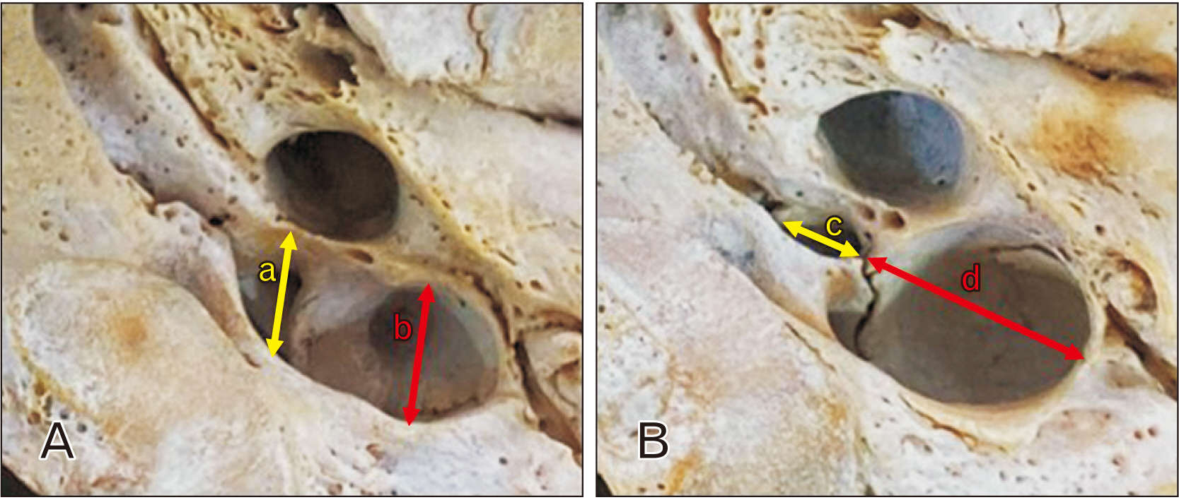
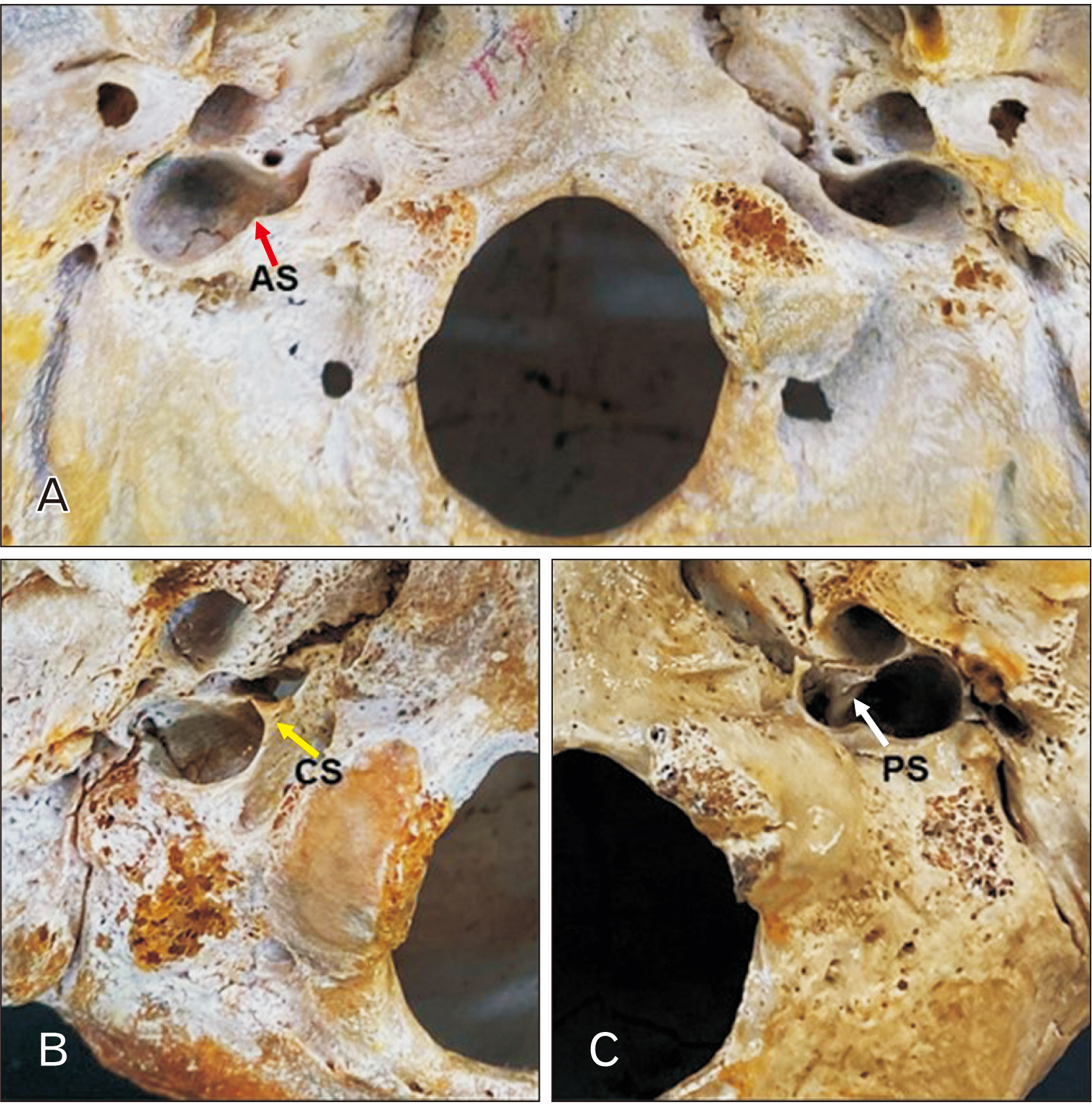
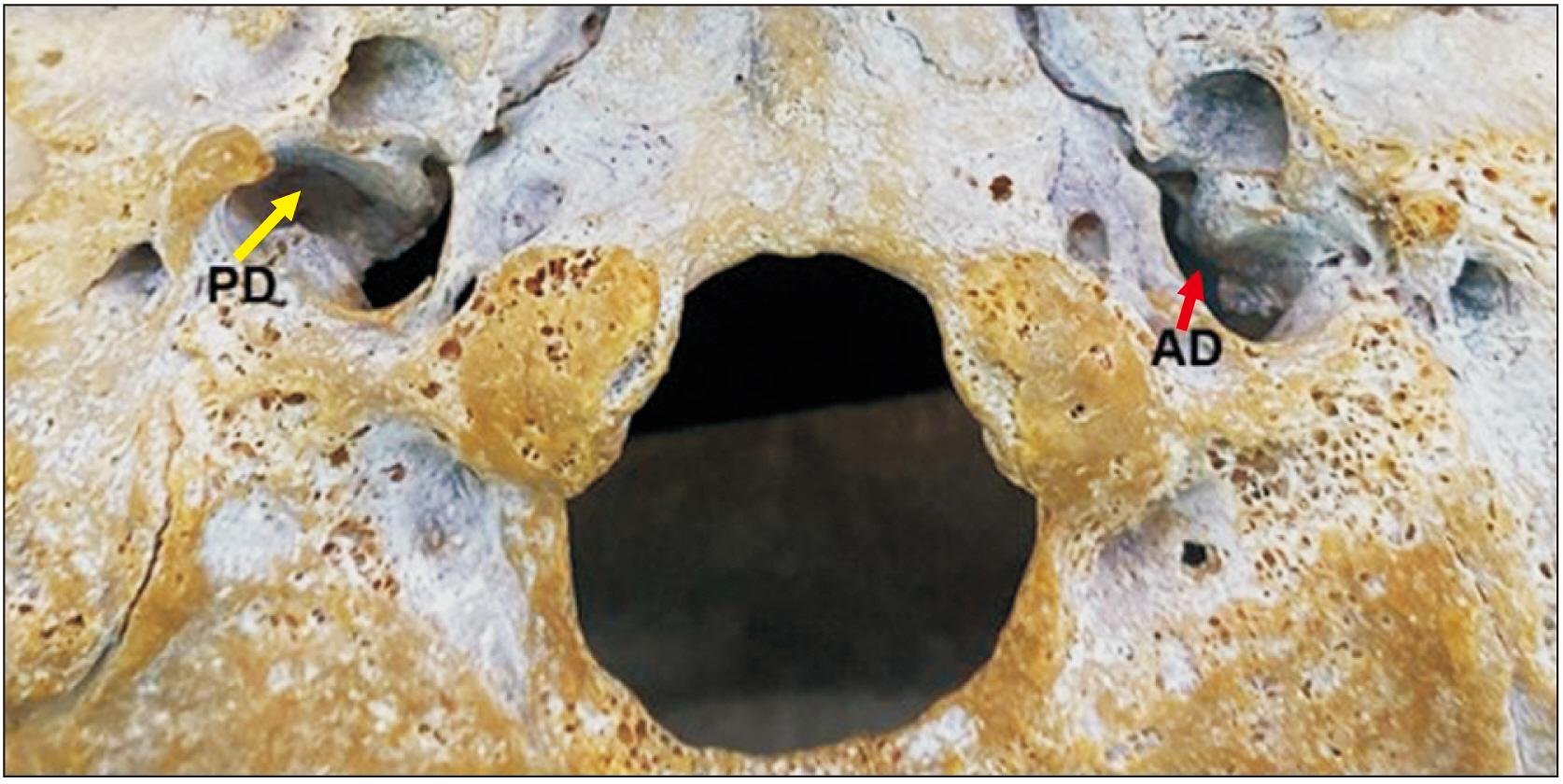
- The jugular foramen (JF) is located between the temporal and occipital bones. The JF is a primary pathway for venous outflow from the skull and passage of nerves. Variations are common in this region and may have clinical and surgical implications. To analyze the sexual dimorphism and JF morphology in skulls from Northeastern Brazil. 128 human skulls from the Anatomy Laboratory of the Federal University of Paraíba, 64 male and 64 female, were selected and the JFs analyzed for bone septation and the presence of a dome. Data analysis considered P<0.05 as significant. On at least one side, complete septation was observed in 26 skulls (20.3%), incomplete septation in 93 skulls (72.6%) and 61 skulls (47.6%) did not present septation. In 114 skulls (89%), 47.6% female and 41.4% male, have a unilateral presence of the dome and 71 (55.4%) have it bilaterally. Posterolateral compartment diameters and JF area had higher values on the right side in the total sample and separated by sex (P<0.05). Most morphometric variables of the anteromedial compartment were higher in male than in female (P<0.05), fact that was not observed in the posterolateral compartment (P>0.05). This study showed a higher prevalence of complete septation in males compared to females. Morphometric analysis presented a peculiar morphology of the JF in this study. These results suggests that the surgical approach to diseases that affect the JF may be peculiar to the studied population, confirming the importance of morphological analysis of the skull base.
Keyword
Figure
Reference
-
References
1. Pereira GAM, Lopes PTC, Santos AMPV, Krebs WD. 2010; Morphometric aspects of the jugular foramen in dry skulls of adult individuals in Southern Brazil. J Morphol Sci. 27:3–5.2. Standring S. Gray's anatomy: the anatomical basis of clinical practice. 40th ed. Churchill Livingstone;2008. p. 423–34.3. Tubbs RS, Griessenauer CJ, Bilal M, Raborn J, Loukas M, Cohen-Gadol AA. 2015; Dural septation on the inner surface of the jugular foramen: an anatomical study. J Neurol Surg B Skull Base. 76:214–7. DOI: 10.1055/s-0034-1543973. PMID: 26225304. PMCID: PMC4433395.
Article4. Bond JD, Zhang M. 2020; Compartmental subdivisions of the jugular foramen: a review of the current models. World Neurosurg. 136:49–57. DOI: 10.1016/j.wneu.2019.12.178. PMID: 31926358.
Article5. Caldeira JVC, Godas AG de L, Carvalho GB de A, Silva KRT da, Machado AR da SR, Almeida PF de, Silva AV da. 2020; Measurement of the jugular foramen in dry skulls from the midwest region of Brazil. Braz J Health Rev. 3:14614–28. Portuguese. DOI: 10.34119/bjhrv3n5-256.6. Thintharua P, Chentanez V. 2023; Morphological analysis and morphometry of the occipital condyle and its relationship to the foramen magnum, jugular foramen, and hypoglossal canal: implications for craniovertebral junction surgery. Anat Cell Biol. 56:61–8. DOI: 10.5115/acb.22.105. PMID: 36635090. PMCID: PMC9989787.
Article7. Shruthi BN, Pavan PH, Shaik SH. 2015; Study on morphological variations in structure of the jugular foramen. Int J Anat Res. 3:1533–35. DOI: 10.16965/ijar.2015.268.
Article8. Jegnie MJ, Asmare Y, Muche A. 2020; Anthropometric measurements of jugular foramen in Ethiopian dried adult skulls. Ethiop J Health Biomed Sci. 10:55–63.9. Shapiro R. 1972; Compartmentation of the jugular foramen. J Neurosurg. 36:340–3. DOI: 10.3171/jns.1972.36.3.0340. PMID: 5059973.
Article10. Das SS, Saluja S, Vasudeva N. 2016; Complete morphometric analysis of jugular foramen and its clinical implications. J Craniovertebr Junction Spine. 7:257–64. DOI: 10.4103/0974-8237.193268. PMID: 27891036. PMCID: PMC5111328.
Article11. Akdag UB, Ogut E, Barut C. 2020; Intraforaminal dural septations of the jugular foramen: a cadaveric study. World Neurosurg. 141:e718–27. DOI: 10.1016/j.wneu.2020.05.271. PMID: 32522647.
Article12. Fang Y, Olewnik Ł, Iwanaga J, Loukas M, Dumont AS, Tubbs RS. 2022; Variations and classification of bony septations of the jugular foramen: an anatomic and histologic study with application to imaging and surgery of the skull base. World Neurosurg. 163:e464–70. DOI: 10.1016/j.wneu.2022.04.010. PMID: 35398325.
Article13. Dichiro G, Fisher RL, Nelson KB. 1964; The jugular foramen. J Neurosurg. 21:447–60. DOI: 10.3171/jns.1964.21.6.0447. PMID: 14171303.
Article14. Keles B, Semaan MT, Fayad JN. 2009; The medial wall of the jugular foramen: a temporal bone anatomic study. Otolaryngol Head Neck Surg. 141:401–7. DOI: 10.1016/j.otohns.2009.05.030. PMID: 19716021.15. Graham MD. 1977; The jugular bulb: its anatomic and clinical considerations in contemporary otology. Laryngoscope. 87:105–25. DOI: 10.1288/00005537-197701000-00013. PMID: 187882.
Article16. Graham MD. 1975; The jugular bulb. Its anatomic and clinical considerations in contemporary otology. Arch Otolaryngol. 101:560–4. DOI: 10.1001/archotol.1975.00780380038010. PMID: 1164239.
Article17. Cömert E, Kiliç C, Cömert A. 2018; Jugular bulb anatomy for lateral skull base approaches. J Craniofac Surg. 29:1969–72. DOI: 10.1097/SCS.0000000000004637. PMID: 29944552.
Article18. Shruthi BN, Pavan PH, Shaik HS, Henjarappa KS. 2015; Morphometric study of jugular foramen. Int J Intg Med Sci. 2:164–6. DOI: 10.18535/ijmsci/v2i8.01. PMID: 25336833. PMCID: PMC4201011.19. Ramina R, Maniglia JJ, Fernandes YB, Paschoal JR, Pfeilsticker LN, Neto MC, Borges G. 2004; Jugular foramen tumors: diagnosis and treatment. Neurosurg Focus. 17:E5. DOI: 10.3171/foc.2004.17.2.5. PMID: 15329020.
Article20. Vanrell JP. Odontologia legal & antropologia forense. Guanabara Koogan;2009.21. Ahmed MM, Jeelani M, Tarnum A. 2015; Anthropometry: a comparative study of right and left sided foramen ovale, jugular foramen and carotid canal. Int J Sci Stud. 3:88–94.22. Gupta C, Kurian P, Seva KN, Kalthur SG, D'Souza AS. 2014; A morphological and morphometric study of jugular foramen in dry skulls with its clinical implications. J Craniovertebr Junction Spine. 5:118–21. DOI: 10.4103/0974-8237.142305. PMID: 25336833. PMCID: PMC4201011.
Article23. Sturrock RR. 1988; Variations in the structure of the jugular foramen of the human skull. J Anat. 160:227–30. PMID: 3253258. PMCID: PMC1262066.24. Chong VF, Fan YF. 1996; Jugular foramen involvement in nasopharyngeal carcinoma. J Laryngol Otol. 110:987–90. DOI: 10.1017/S0022215100135534. PMID: 8977870.
Article25. Wysocki J, Chmielik LP, Gacek W. 1999; Variability of magnitude of the human jugular foramen in relation to conditions of the venous outflow after ligation of the internal jugular vein. Otolaryngol Pol. 53:173–7. Polish.26. Padget DH. Contributions to embryology. Vol. 36, The development of the cranial venous system in man: from the viewpoint of comparative anatomy. Carnegie Institution of Washington Publication;1957. p. 79–140.27. Vlajković S, Vasović L, Daković-Bjelaković M, Stanković S, Popović J, Cukuranović R. 2010; Human bony jugular foramen: some additional morphological and morphometric features. Med Sci Monit. 16:BR140–6. PMID: 20424543.28. Vijisha P, Bilodi AK, Anandaraj L. 2013; Morphometric study of jugular foramen in Tamil Nadu region. Natl J Clin Anat. 2:71–4. DOI: 10.4103/2277-4025.297875.
Article
- Full Text Links
- Actions
-
Cited
- CITED
-
- Close
- Share
- Similar articles
-
- Morphometric analysis of infraorbital foramen in Indian dry skulls
- Jugular foramen neurilemmoma mimicking an intra-axial brainstem tumor: a case report
- The study of hyperostosic variants: significance of hyperostotic variants of human skulls in anthropology
- Morphological analysis and morphometry of the occipital condyle and its relationship to the foramen magnum, jugular foramen, and hypoglossal canal: implications for craniovertebral junction surgery
- Morphological study of styloid process of the temporal bone and its clinical implications


