Anat Cell Biol.
2024 Jun;57(2):204-212. 10.5115/acb.23.293.
Anatomical study of the zygomaticofacial foramen and zygomatic canals communicating with the zygomaticofacial foramen for zygomatic implant treatment: a cadaver study with micro-computed tomography analysis
- Affiliations
-
- 1Department of Anatomy, The Nippon Dental University School of Life Dentistry at Tokyo, Tokyo, Japan
- KMID: 2556564
- DOI: http://doi.org/10.5115/acb.23.293
Abstract
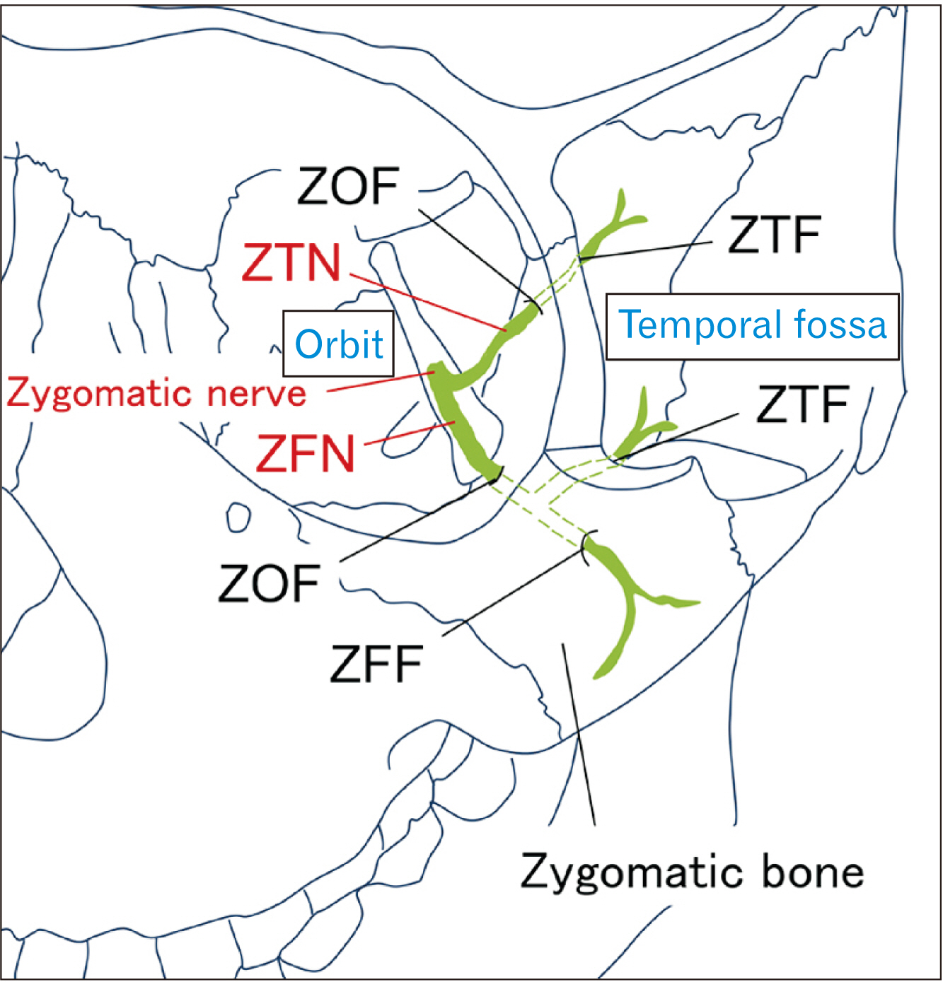
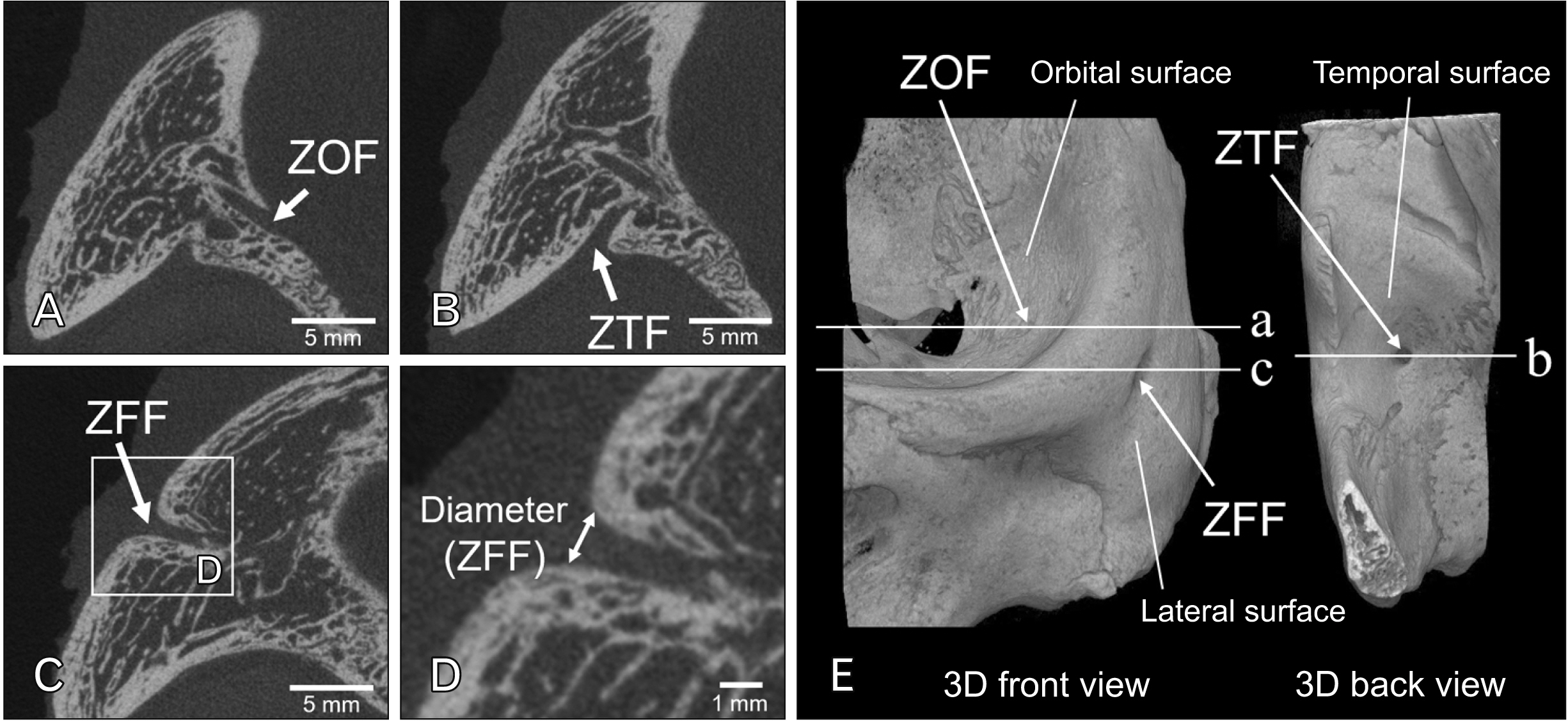
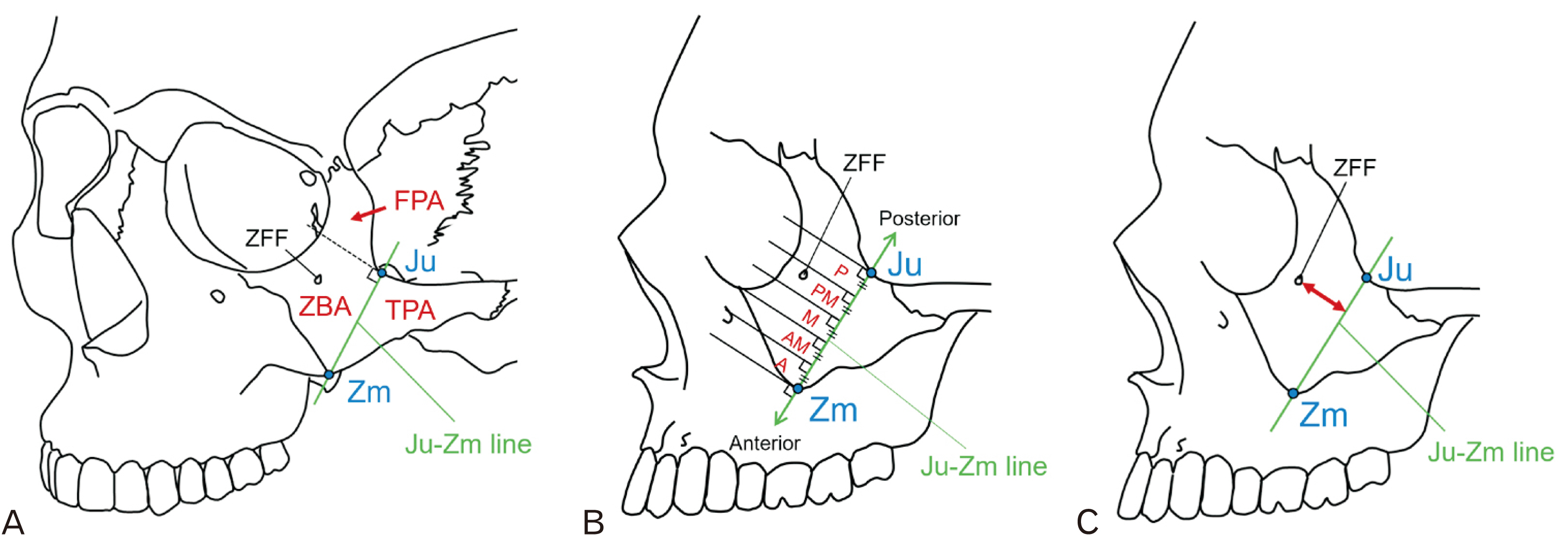
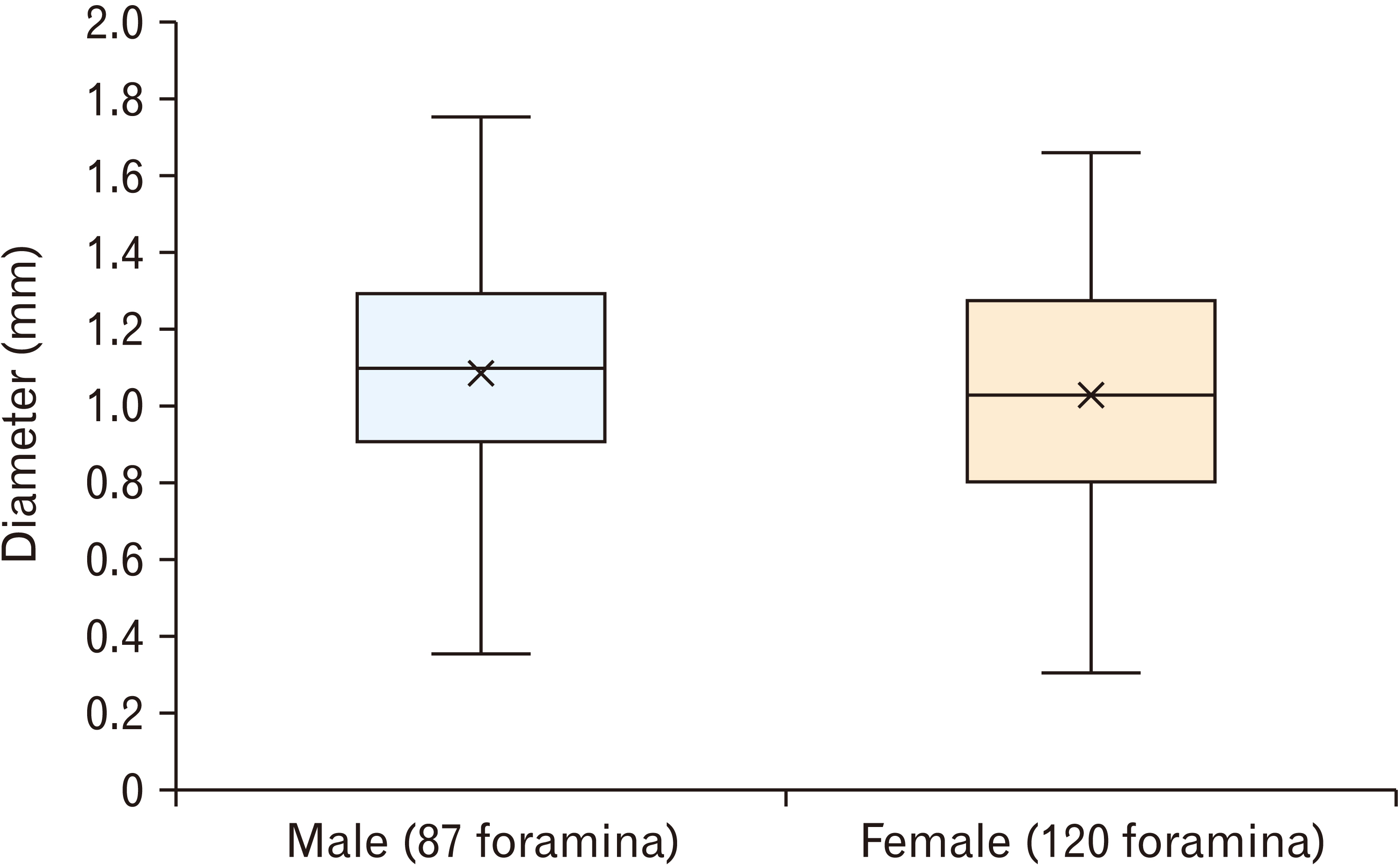
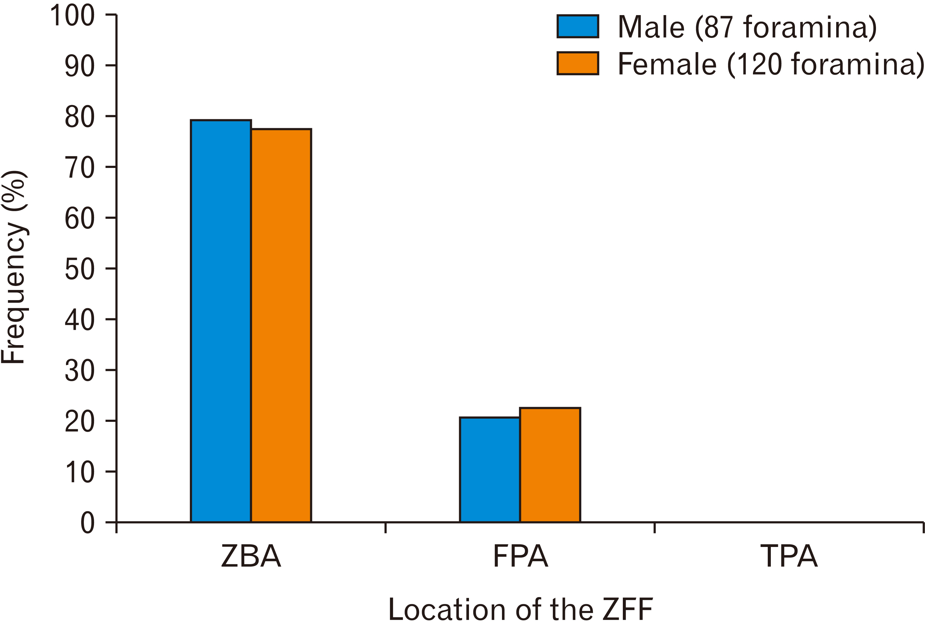
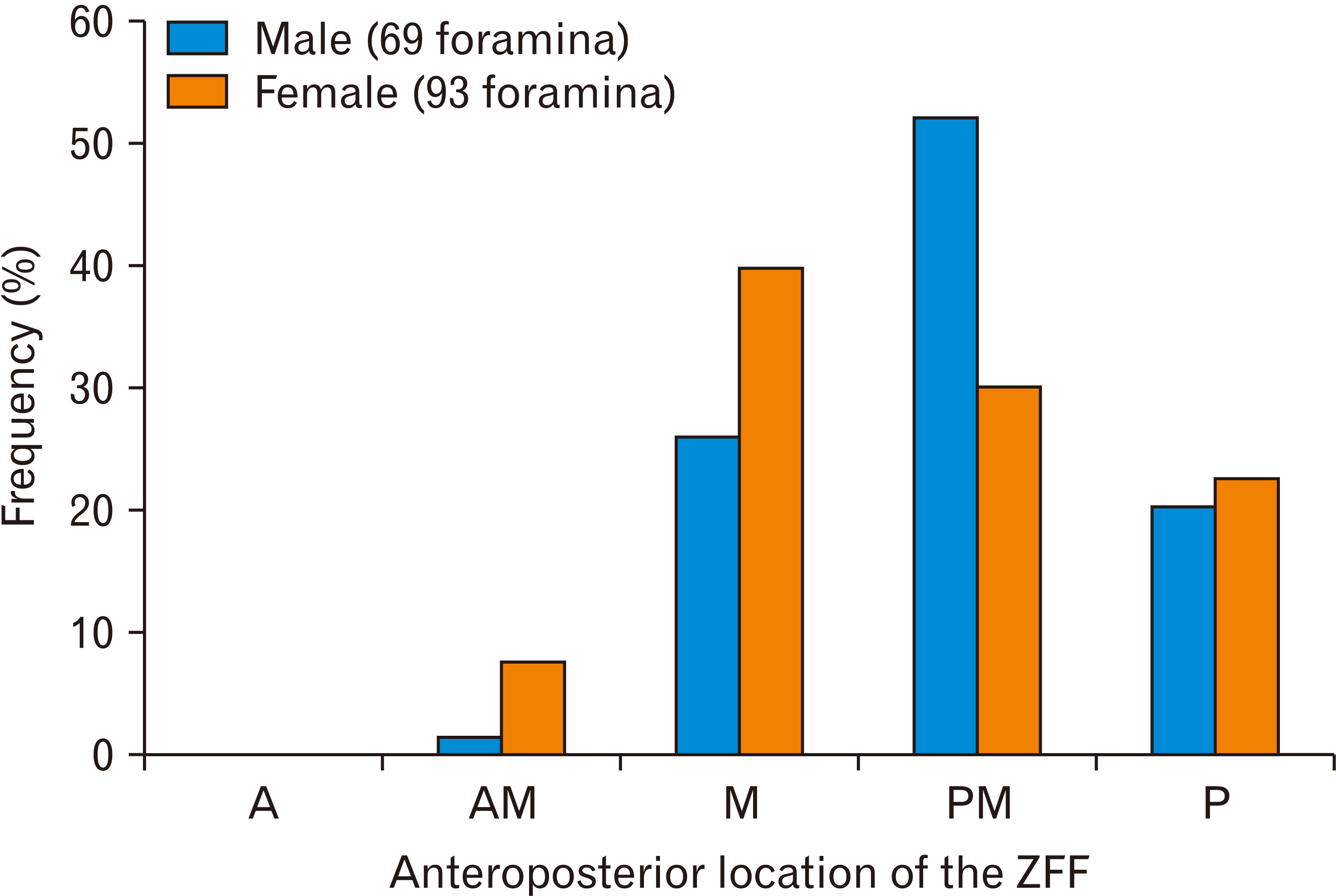
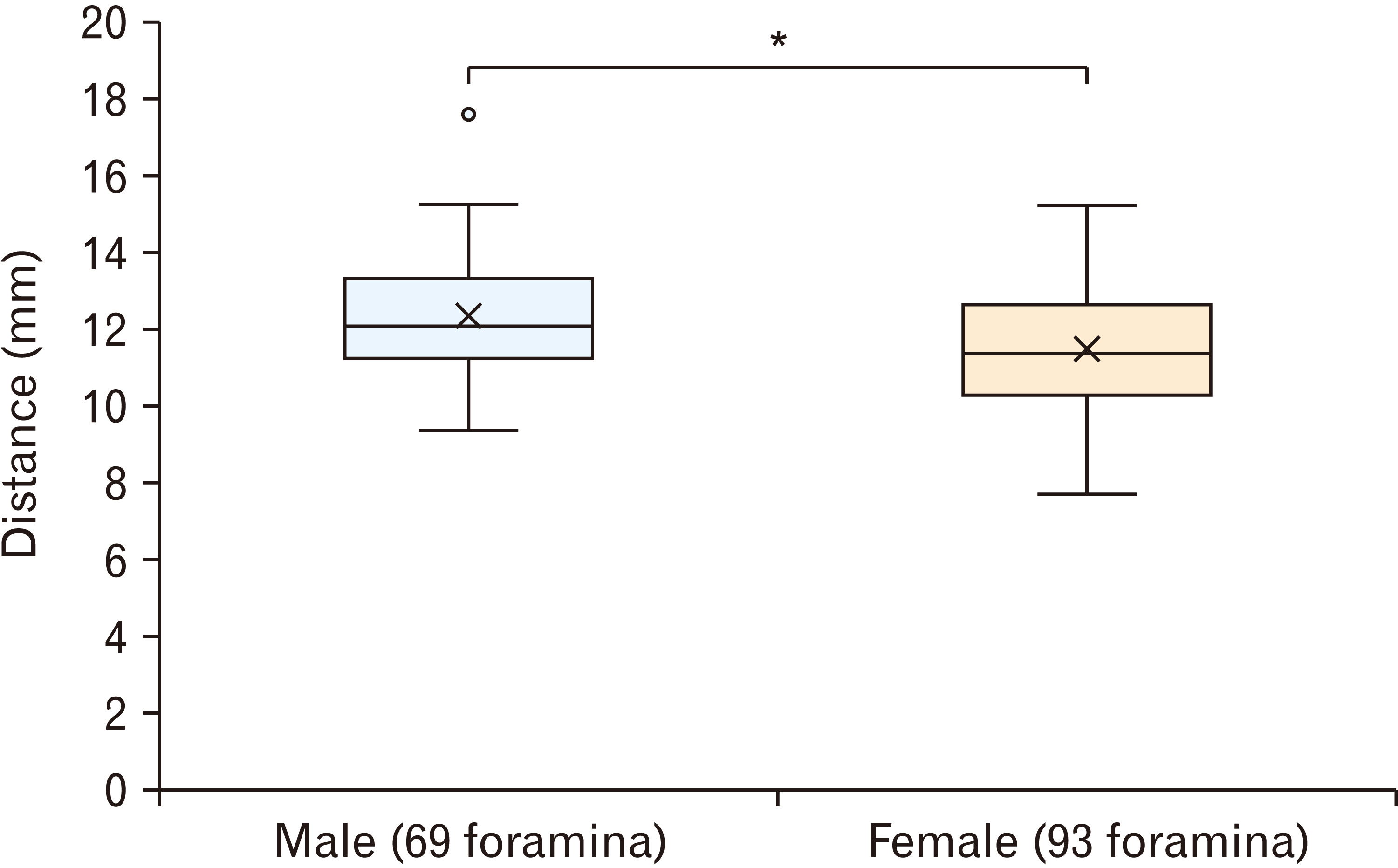
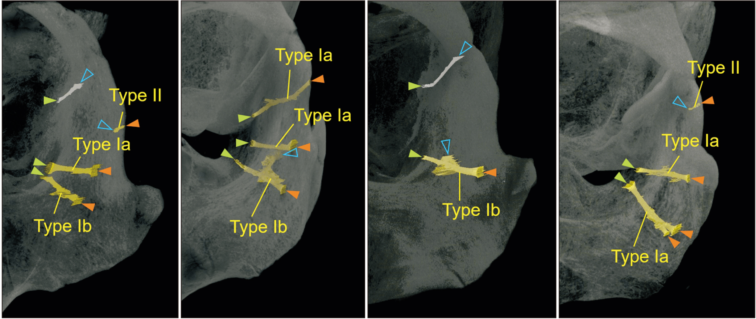
- In the present study, anatomical assessment of zygomaticofacial foramina (ZFFs) and zygomatic canals communicating with ZFFs were performed using cadaver micro-computed tomography images. It was suggested that all ZFFs were located above the jugale (Ju)-zygomaxillare (Zm) line, which is the reference line connecting the Ju and Zm, and most were located in the zygomatic body area (ZBA). The anteroposterior position of the ZFF in the ZBA was within a middle to posterior region and was most often located slightly posteriorly in males and closer to the middle of the region in females. The mean distance from the Ju-Zm line to the ZFF in the ZBA was 12.36 mm (standard deviation [SD] 1.52 mm) in males and 11.48 mm (SD 1.61 mm) in females. In zygomatic canals communicating with ZFFs, most zygomatic canals were type I canals, communicating from the zygomaticoorbital foramen and harboring the zygomaticofacial nerve, and the others were type II canals, communicating from the zygomaticotemporal foramen and located near the posterior margin of the frontal process. These results provide useful anatomical information for preventing nerve injury during surgical procedures for zygomatic implant treatment.
Keyword
Figure
Reference
-
References
1. Brånemark PI, Gröndahl K, Worthington P. Osseointegration and autogenous onlay bone grafts: reconstruction of the edentulous atrophic maxilla. Quintessence Publishing Co;2001. p. 112–34. DOI: 10.56373/2002-3-24.2. Brånemark PI. The zygomaticus fixture: clinical procedures. Nobel Biocare;1998.3. Bedrossian E, Stumpel L 3rd, Beckely ML, Indresano T. 2002; The zygomatic implant: preliminary data on treatment of severely resorbed maxillae. A clinical report. Int J Oral Maxillofac Implants. 17:861–5. Erratum in: Int J Oral Maxillofac Implants 2003;18:292.4. Malevez C, Daelemans P, Adriaenssens P, Durdu F. 2003; Use of zygomatic implants to deal with resorbed posterior maxillae. Periodontol 2000. 33:82–9. DOI: 10.1046/j.0906-6713.2002.03307.x. PMID: 12950843.
Article5. Malevez C, Abarca M, Durdu F, Daelemans P. 2004; Clinical outcome of 103 consecutive zygomatic implants: a 6-48 months follow-up study. Clin Oral Implants Res. 15:18–22. DOI: 10.1046/j.1600-0501.2003.00985.x. PMID: 15005100.
Article6. Stella JP, Warner MR. 2000; Sinus slot technique for simplification and improved orientation of zygomaticus dental implants: a technical note. Int J Oral Maxillofac Implants. 15:889–93. PMID: 11151591.7. Aparicio C, Alandez J. Zygomatic implants: the anatomy-guided approach. Quintessence Publishing Co.;2012. p. 90–110. p. 2458. Bedrossian E, Bedrossian EA. 2018; Prevention and the management of complications using the Zygoma implant: a review and clinical experiences. Int J Oral Maxillofac Implants. 33:e135–45. DOI: 10.11607/jomi.6539. PMID: 30231096.
Article9. Kahnberg KE, Henry PJ, Hirsch JM, Ohrnell LO, Andreasson L, Brånemark PI, Chiapasco M, Gynther G, Finne K, Higuchi KW, Isaksson S, Malevez C, Neukam FW, Sevetz E Jr, Urgell JP, Widmark G, Bolind P. 2007; Clinical evaluation of the zygoma implant: 3-year follow-up at 16 clinics. J Oral Maxillofac Surg. 65:2033–8. DOI: 10.1016/j.joms.2007.05.013. PMID: 17884535.
Article10. Chrcanovic BR, Abreu MH. 2013; Survival and complications of zygomatic implants: a systematic review. Oral Maxillofac Surg. 17:81–93. DOI: 10.1007/s10006-012-0331-z. PMID: 22562293.
Article11. Bedrossian E. 2010; Rehabilitation of the edentulous maxilla with the zygoma concept: a 7-year prospective study. Int J Oral Maxillofac Implants. 25:1213–21. PMID: 21197500.12. Reichert TE, Kunkel M, Wahlmann U, Wagner W. 1999; The zygomatic implant indications and first clinical experiences. Z Zahnarztl Implantol. 15:65–70.13. Standring S. Gray's anatomy: the anatomical basis of clinical practice. 40th ed. Elsevier/Churchill Livingstone;2008. p. 493–4.14. Hollinshead WH. Anatomy for surgeons. 3rd ed. Harper & Row;1982. p. 146–318.15. Wolff E, Last RJ. Anatomy of the eye and orbit. 5th ed. Saunders Company;1961. p. 6–10. p. 285–6.16. Loukas M, Owens DG, Tubbs RS, Spentzouris G, Elochukwu A, Jordan R. 2008; Zygomaticofacial, zygomaticoorbital and zygomaticotemporal foramina: anatomical study. Anat Sci Int. 83:77–82. DOI: 10.1111/j.1447-073X.2007.00207.x. PMID: 18507616.
Article17. Aksu F, Ceri NG, Arman C, Zeybek FG, Tetik S. 2009; Location and incidence of the zygomaticofacial foramen: an anatomic study. Clin Anat. 22:559–62. DOI: 10.1002/ca.20805. PMID: 19418451.
Article18. Hwang SH, Jin S, Hwang K. 2007; Location of the zygomaticofacial foramen related to malar reduction. J Craniofac Surg. 18:872–4. DOI: 10.1097/scs.0b013e3180a03353. PMID: 17667680.
Article19. Melchenko SA, Cherekaev VA, Alyoshkina OY, Danilov GV, Musa G, Strunina UV, Golbin DA, Lasunin NV, Zaychenko AA. 2022; Assessing the reliability of zygomatic bone landmarks as guides to reach the inferior orbital fissure in orbitozygomatic osteotomy: anatomical study of 83 human skulls. Neurosurg Rev. 45:2175–82. DOI: 10.1007/s10143-021-01726-8. PMID: 35028786.
Article20. Gupta T, Gupta SK. 2009; The ZMF: Is it a reliable intraoperative guide for the IOF? Clin Anat. 22:451–5. DOI: 10.1002/ca.20783. PMID: 19291758.
Article21. Ferro A, Basyuni S, Brassett C, Santhanam V. 2017; Study of anatomical variations of the zygomaticofacial foramen and calculation of reliable reference points for operation. Br J Oral Maxillofac Surg. 55:1035–41. DOI: 10.1016/j.bjoms.2017.10.016. PMID: 29122337.
Article22. Martins C, Li X, Rhoton AL Jr. 2003; Role of the zygomaticofacial foramen in the orbitozygomatic craniotomy: anatomic report. Neurosurgery. 53:168–72. discussion 172–3. DOI: 10.1227/01.NEU.0000068841.17293.BB. PMID: 12823886.
Article23. Zhao Y, Chundury RV, Blandford AD, Perry JD. 2018; Anatomical description of zygomatic foramina in African American skulls. Ophthalmic Plast Reconstr Surg. 34:168–71. DOI: 10.1097/IOP.0000000000000905. PMID: 28369018. PMCID: PMC5623600.
Article24. Deana NF, Alves N. 2020; Frequency and location of the zygomaticofacial foramen and its clinical importance in the placement of zygomatic implants. Surg Radiol Anat. 42:823–30. DOI: 10.1007/s00276-020-02455-1. PMID: 32246188.
Article25. Mangal A, Choudhry R, Tuli A, Choudhry S, Choudhry R, Khera V. 2004; Incidence and morphological study of zygomaticofacial and zygomatico-orbital foramina in dry adult human skulls: the non-metrical variants. Surg Radiol Anat. 26:96–9. DOI: 10.1007/s00276-003-0198-7. PMID: 15004726.
Article26. Iwanaga J, Badaloni F, Watanabe K, Yamaki KI, Oskouian RJ, Tubbs RS. 2018; Anatomical study of the zygomaticofacial foramen and its related canal. J Craniofac Surg. 29:1363–5. DOI: 10.1097/SCS.0000000000004457. PMID: 29521755.
Article27. Kim HS, Oh JH, Choi DY, Lee JG, Choi JH, Hu KS, Kim HJ, Yang HM. 2013; Three-dimensional courses of zygomaticofacial and zygomaticotemporal canals using micro-computed tomography in Korean. J Craniofac Surg. 24:1565–8. DOI: 10.1097/SCS.0b013e318299775d. PMID: 24036727.
Article28. Kato Y, Kizu Y, Tonogi M, Ide Y, Yamane GY. 2005; Internal structure of zygomatic bone related to zygomatic fixture. J Oral Maxillofac Surg. 63:1325–9. DOI: 10.1016/j.joms.2005.05.313. PMID: 16122597.
Article29. Ide Y, Nakahara T, Nasu M, Matsunaga S, Iwanaga T, Tominaga N, Tamaki Y. 2013; Postnatal mandibular cheek tooth development in the miniature pig based on two-dimensional and three-dimensional X-ray analyses. Anat Rec (Hoboken). 296:1247–54. DOI: 10.1002/ar.22725. PMID: 23749549.
Article30. von Arx T, Lozanoff S, Bosshardt D. 2014; Accessory mental foramina. Oral Surg. 7:216–27. DOI: 10.1111/ors.12071. PMID: 27146294.31. Pelé A, Berry PA, Evanno C, Jordana F. 2021; Evaluation of mental foramen with cone beam computed tomography: a systematic review of literature. Radiol Res Pract. 2021:8897275. DOI: 10.1155/2021/8897275. PMID: 33505723. PMCID: PMC7806401.
Article32. Gümüşok M, Akarslan ZZ, Başman A, Üçok Ö. 2017; Evaluation of accessory mental foramina morphology with cone-beam computed tomography. Niger J Clin Pract. 20:1550–4. DOI: 10.4103/1119-3077.187329. PMID: 29378985.
Article33. Gungor E, Aglarci OS, Unal M, Dogan MS, Guven S. 2017; Evaluation of mental foramen location in the 10-70 years age range using cone-beam computed tomography. Niger J Clin Pract. 20:88–92. DOI: 10.4103/1119-3077.178915. PMID: 27958253.
Article34. Kalender A, Orhan K, Aksoy U. 2012; Evaluation of the mental foramen and accessory mental foramen in Turkish patients using cone-beam computed tomography images reconstructed from a volumetric rendering program. Clin Anat. 25:584–92. DOI: 10.1002/ca.21277. PMID: 21976294.
Article35. Hong JH, Kim HJ, Hong JH, Park KB. 2022; Study of infraorbital foramen using 3-dimensional facial bone computed tomography scans. Pain Physician. 25:E127–32. PMID: 35051160.36. Désiré A, Ebogo M, Amougou M, Essono N, Zogo O. 2023; Assessment of infraorbital foramen position using computed tomography-scan in a cohort of Cameroonian adults: landmarks in facial surgery and anesthesiology. Pan Afr Med J. 45:134. DOI: 10.11604/pamj.2023.45.134.37733. PMID: 37790162. PMCID: PMC10543902.37. Dagistan S, Miloǧlu Ö, Altun O, Umar EK. 2017; Retrospective morphometric analysis of the infraorbital foramen with cone beam computed tomography. Niger J Clin Pract. 20:1053–64. DOI: 10.4103/1119-3077.217247. PMID: 29072226.
Article38. Siddiqui HF, Konschake M, Ottone NE, Olewnik Ł, Iwanaga J, Aysenne A, Xu L, Tubbs RS. 2023; A marginal process of the zygomatic bone predicts a lateral exit of the zygomaticotemporal nerve: an anatomical study with application to surgery around the midface. Clin Anat. 36:708–14. DOI: 10.1002/ca.24021. PMID: 36752958.
Article
- Full Text Links
- Actions
-
Cited
- CITED
-
- Close
- Share
- Similar articles
-
- Measurements of the Zygomatic Bones and Morphology of the Zygomaticofacial and Zygomaticotemporal foramina in Korean
- A Morphometric Analysis of the Foramen Ovale and the Zygomatic Points Determined by a Computed Tomography in Patients with Idiopathic Trigeminal Neuralgia
- Non-Metrical Morphologic Variations of Korean Skull Foramina
- Anatomical structure of lingual foramen in cone beam computed tomography
- A giant foramen of Vesalius: case report

