Anat Cell Biol.
2024 Jun;57(2):183-193. 10.5115/acb.24.027.
Anatomy of the clitoris: the corona of the glans clitoris, clitoral coronal papillae, and the coronopreputial frenulum
- Affiliations
-
- 1Department of Pathology, Anatomy, and Laboratory Medicine (PALM), West Virginia University School of Medicine, Morgantown, WV, USA
- KMID: 2556562
- DOI: http://doi.org/10.5115/acb.24.027
Abstract
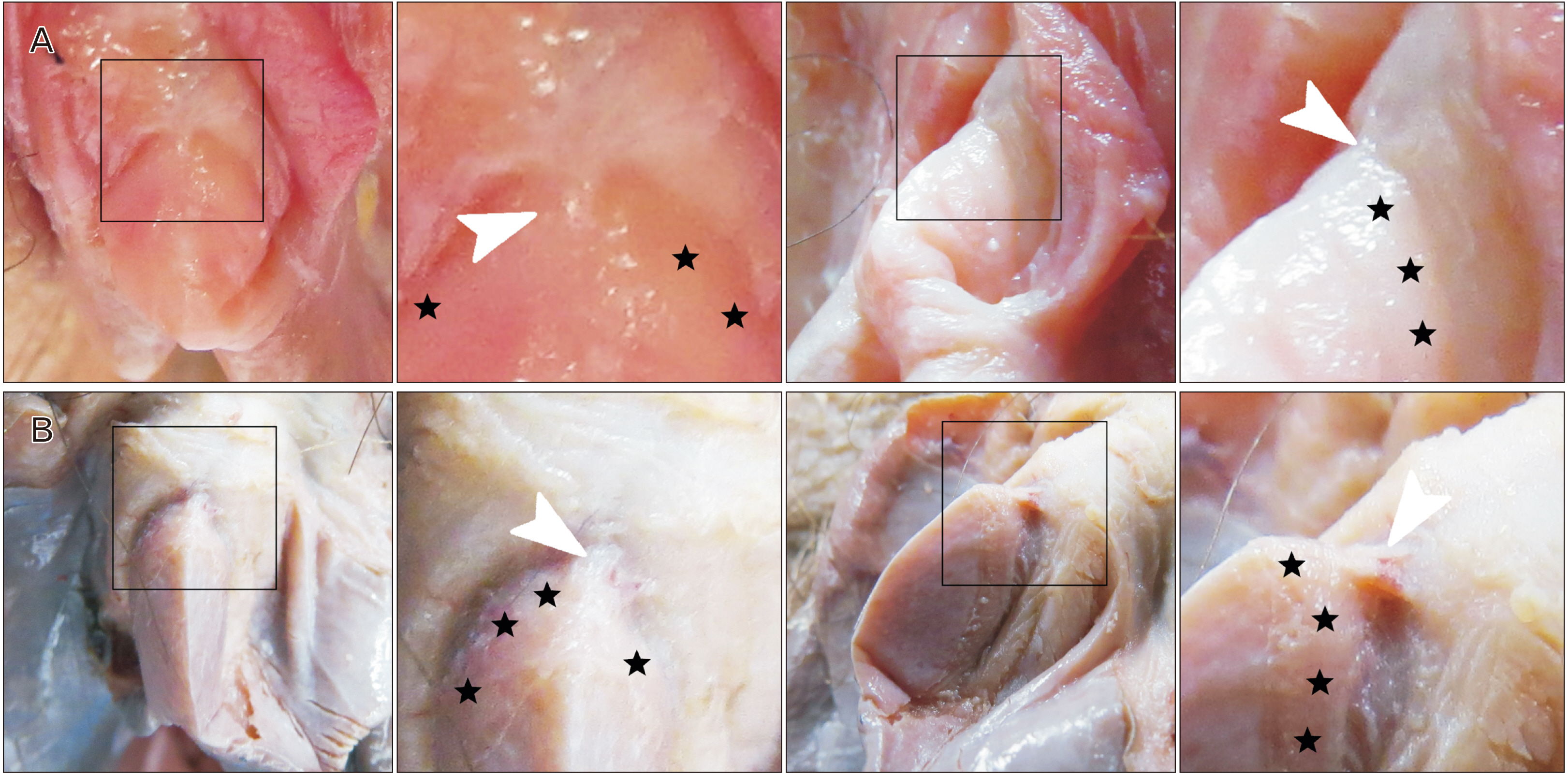
- The corona of the glans clitoris is a clinically important yet poorly understood anatomical structure. There has been longstanding confusion regarding the prevalence of the corona of the glans clitoris and, moreover, its very existence. Therefore, this anatomical study assesses the prevalence of the corona of the glans clitoris and the gross anatomy of the proximal glans clitoris. Anatomy was assessed in 104 female donor bodies ranging in age from 50 to 102 years with an average age-at-death of 78.1±10.9 years (mean±SD). All clitorises (100%; 104:104 dorsums and 100%; 208:208 sides) were found to have a well-defined clitoral corona. Three of 104 (2.9%) coronas possessed grossly visible, outward-projecting, bluntly rounded papillae. Some donors possessed a coronopreputial frenulum. Clitoropreputial adhesions were common and associated with clitoral pearls. Clitoral pearls were identified in 37.8% (14:37) of unembalmed donors and observed to create clitoral craters, structural deformations in the surface of the corona and glans. The results of this study suggest that the corona of the glans clitoris is a ubiquitous anatomical structure. The clitoral coronal papillae and coronopreputial frenulum are novel, previously undescribed, anatomical structures. This study identifies that the corona of the glans clitoris is prone to pathological processes such as clitoral pearl formation and clitoral deformation. In addition to novel anatomical findings, the results of this study call attention to the need for life-long clitoral examinations. Furthermore, the corona of the glans clitoris should be regularly included in anatomical texts and accurately depicted in anatomical illustrations.
Keyword
Figure
Reference
-
1. Allen LE, Hardy BE, Churchill BM. 1982; The surgical management of the enlarged clitoris. J Urol. 128:351–4. DOI: 10.1016/S0022-5347(17)52923-7. PMID: 7109108.
Article2. Farkas A, Chertin B, Hadas-Halpren I. 2001; 1-Stage feminizing genitoplasty: 8 years of experience with 49 cases. J Urol. 165(6 Pt 2):2341–6. DOI: 10.1016/S0022-5347(05)66199-X. PMID: 11371974.
Article3. Perovic SV, Djordjevic ML. 2003; Metoidioplasty: a variant of phalloplasty in female transsexuals. BJU Int. 92:981–5. DOI: 10.1111/j.1464-410X.2003.04524.x. PMID: 14632860.
Article4. Lesma A, Bocciardi A, Montorsi F, Rigatti P. 2007; Passerini-glazel feminizing genitoplasty: modifications in 17 years of experience with 82 cases. Eur Urol. 52:1638–44. DOI: 10.1016/j.eururo.2007.02.068. PMID: 17408848.
Article5. Kujur AR, Joseph V, Chandra P. 2016; Nerve sparing clitoroplasty in a rare case of idiopathic clitoromegaly. Indian J Plast Surg. 49:86–90. DOI: 10.4103/0970-0358.182241. PMID: 27274128. PMCID: PMC4878251.
Article6. Adams MC, DeMarco RT, Thomas JC. Docimo SG, Canning D, Khoury A, Salle JLP, editors. Surgery for intersex disorders and urogenital sinus. The Kelalis-King-Belman textbook of clinical pediatric urology. 6th ed. CRC Press;2018. p. 1215–31.7. Aerts L, Rubin RS, Randazzo M, Goldstein SW, Goldstein I. 2018; Retrospective study of the prevalence and risk factors of clitoral adhesions: women's health providers should routinely examine the glans clitoris. Sex Med. 6:115–22. DOI: 10.1016/j.esxm.2018.01.003. PMID: 29559206. PMCID: PMC5960030.
Article8. Myers MC, Romanello JP, Nico E, Marantidis J, Rowen TS, Sussman RD, Rubin RS. 2022; A retrospective case series on patient satisfaction and efficacy of non-surgical lysis of clitoral adhesions. J Sex Med. 19:1412–20. DOI: 10.1016/j.jsxm.2022.06.011. PMID: 35869023.
Article9. Verkauf BS, Von Thron J, O'Brien WF. 1992; Clitoral size in normal women. Obstet Gynecol. 80:41–4. PMID: 1603495.10. Nigam A, Prakash A, Saxena P, Yadav R, Raghunandan C. 2011; Hirsutism and abnormal genitalia. J Indian Acad Clin Med. 12:46–8.11. Mayer JCA. Beschreibung des ganzen menschlichen körpers, mit den wichtigsten neueren anatomischen entdeckungen bereichert, nebst physiologischen erläuterungen. G. J. Decker. 1788. German.12. Bernstein JG. Handbuch nach alphabetischer ordnung über die vorzüglichsten gegenstände der anatomie, physiologie und gerichtliche arzneigelahrtheit, für praktische wundärzte. Schwickertschen Verlage. 1794. German.13. Hempel AF. Anfangsgründe der anatomie. Johann Christian Daniel Schneider. 1801. German.14. Moser A. Lehrbuch der geschlechtskrankheiten des weibes, nebst einem anhange, enthaltend die regeln für die untersuchung der weiblichen geschlechtstheile. Nach den neuesten quellen und eigener erfahrung. Hirschwald. 1843. German.15. Eckley WT, Eckley CB. Practical anatomy, including a special section on the fundamental principles of anatomy. P. Blakiston's Son & Co.;1899. DOI: 10.5962/bhl.title.29837.16. Eckley WT, Eckley CB. A manual of dissection and practical anatomy: founded on Gray and Gerrish. Lea Brothers & Company;1903. DOI: 10.1097/00000441-190305000-00023.17. Pratt EH. 1914; Sympathetic nerve waste. JAAOS. 2:62–6.18. Di Marino V, Lepidi H. Di Marino V, Lepidi H, editors. External morphology of the clitoris. Anatomic study of the clitoris and the bulbo-clitoral organ. Springer;2014. p. 27–38. DOI: 10.1007/978-3-319-04894-9_4.19. Federative International Programme for Anatomical Terminology. Terminologia anatomica. 2nd ed. International Anatomical Terminology;2019.20. Jeppson PC, Balgobin S, Washington BB, Hill AJ, Lewicky-Gaupp C, Wheeler T 2nd, Ridgeway B, Mazloomdoost D, Balk EM, Corton MM, DeLancey J. Society of Gynecologic Surgeons Pelvic Anatomy Group. 2018; Recommended standardized terminology of the anterior female pelvis based on a structured medical literature review. Am J Obstet Gynecol. 219:26–39. DOI: 10.1016/j.ajog.2018.04.006. PMID: 29630884.
Article21. Hill AJ, Balgobin S, Mishra K, Jeppson PC, Wheeler T 2nd, Mazloomdoost D, Anand M, Ninivaggio C, Hamner J, Bochenska K, Mama ST, Balk EM, Corton MM, Delancey J. Society of Gynecologic Surgeons Pelvic Anatomy Group. 2021; Recommended standardized anatomic terminology of the posterior female pelvis and vulva based on a structured medical literature review. Am J Obstet Gynecol. 225:169.e1–16. DOI: 10.1016/j.ajog.2021.02.033. PMID: 33705749.
Article22. Zdilla MJ. 2022; Recommended standardized anatomic terminology of the posterior female pelvis and vulva. Am J Obstet Gynecol. 227:118. DOI: 10.1016/j.ajog.2022.02.018. PMID: 35183502.
Article23. Zdilla MJ. 2022; What is a vulva? Anat Sci Int. 97:323–46. DOI: 10.1007/s12565-022-00674-7. PMID: 35704265.
Article24. Hill AJ, Balgobin S, Delancey J. 2022; Reply: Further discussion and clarification of female vulvar anatomy. Am J Obstet Gynecol. 227:119. DOI: 10.1016/j.ajog.2022.02.017. PMID: 35183501.
Article25. Goldstein I, Meston CM, Davis S, Traish A. Women's sexual function and dysfunction: study, diagnosis and treatment. Taylor & Francis;2005. DOI: 10.1201/b14618.26. Khatun S. Principles and practices of premalignant and malignant disorders of vulva. Jaypee Brothers Medical Publishers;2019.27. Fahmy MAB. Normal and abnormal prepuce. Springer;2020. DOI: 10.1007/978-3-030-37621-5.28. Radke SM, Stockdale CK. Goldstein AT, Pukall CF, Goldstein I, Krapf JM, Goldstein SW, Goldstein G, editors. Chronic clitoral pain and clitorodynia. Female sexual pain disorders: evaluation and management. 2nd ed. John Wiley & Sons;2020. p. 375–80. DOI: 10.1002/9781119482598.ch41.29. Sadeghipour Roudsari S, Esmailzadehha N. 2010; Aposthia: a case report. J Pediatr Surg. 45:e17–9. DOI: 10.1016/j.jpedsurg.2010.05.030. PMID: 20713198.
Article30. Nayak SB, Pai SU, Shenoy MG, Reghunathan D. 2018; Accessory fold of skin on the ventral surface of the penis: is it a redundant prepuce? Andrologia. 50:e12941. DOI: 10.1111/and.12941. PMID: 29315704.
Article31. Esse I, Kincaid CM, Terrell CA, Mesinkovska NA. 2023; Female genital mutilation: overview and dermatologic relevance. JAAD Int. 14:92–8. DOI: 10.1016/j.jdin.2023.07.022. PMID: 38352964. PMCID: PMC10862004.
Article32. Deibert GA. 1933; The separation of the prepuce in the human penis. Anat Rec. 57:387–99. DOI: 10.1002/ar.1090570409.
Article33. Gairdner D. 1949; The fate of the foreskin, a study of circumcision. Br Med J. 2:1433–7. illust. DOI: 10.1136/bmj.2.4642.1433. PMID: 15408299. PMCID: PMC2051968.34. Sonthalia S, Singal A. 2016; Smegma pearls in young uncircumcised boys. Pediatr Dermatol. 33:e186–9. DOI: 10.1111/pde.12832. PMID: 27071486.
Article35. Baskin L, Shen J, Sinclair A, Cao M, Liu X, Liu G, Isaacson D, Overland M, Li Y, Cunha GR. 2018; Development of the human penis and clitoris. Differentiation. 103:74–85. DOI: 10.1016/j.diff.2018.08.001. PMID: 30249413. PMCID: PMC6234061.
Article36. Liu X, Liu G, Shen J, Yue A, Isaacson D, Sinclair A, Cao M, Liaw A, Cunha GR, Baskin L. 2018; Human glans and preputial development. Differentiation. 103:86–99. DOI: 10.1016/j.diff.2018.08.002. PMID: 30245194. PMCID: PMC6234068.
Article37. Puri A, Sikdar S, Prakash R. 2017; Pediatric penile and glans anthropometry nomograms: an aid in hypospadias management. J Indian Assoc Pediatr Surg. 22:9–12. DOI: 10.4103/0971-9261.194610. PMID: 28082769. PMCID: PMC5217151.
Article38. van der Putte SC, Sie-Go DM. 2011; Development and structure of the glandopreputial sulcus of the human clitoris with a special reference to glandopreputial glands. Anat Rec (Hoboken). 294:156–64. DOI: 10.1002/ar.21279. PMID: 21157926.
Article39. Krapf J, Mautz T, Lorenzini S, Holloway J, Goldstein A. 2022; Clinical presentation of clitorodynia associated with clitoral adhesions and keratin pearls. J Sex Med. 19(8 Suppl 3):S14. DOI: 10.1016/j.jsxm.2022.05.032.
Article40. Bragiel RM, Umasankar N, Burgis JT, Tomlin KV. 2023; Treatment of clitoral keratin pearls with topical estrogen cream: case report. J Pediatr Adolesc Gynecol. 36:321–3. DOI: 10.1016/j.jpag.2022.10.002. PMID: 36209998.
Article41. Winfrey OK, Fei YF, Dendrinos ML, Rosen MW, Smith YR, Quint EH. 2022; Lichen sclerosus throughout childhood and adolescence: not only a premenarchal disease. J Pediatr Adolesc Gynecol. 35:624–8. DOI: 10.1016/j.jpag.2022.08.011. PMID: 36038010.
Article42. Winter AG, Rubin RS. Goldstein AT, Pukall CF, Goldstein I, Krapf JM, Goldstein SW, Goldstein G, editors. Vulvoscopic evaluation of vulvodynia. Female sexual pain disorders: evaluation and management. 2nd ed. John Wiley & Sons;2020. p. 125–31. DOI: 10.1002/9781119482598.ch15.43. Romanello JP, Myers MC, Nico E, Rubin RS. 2023; Clitoral adhesions: a review of the literature. Sex Med Rev. 11:196–201. DOI: 10.1093/sxmrev/qead004. PMID: 36973166.
Article44. Tagatz GE, Kopher RA, Nagel TC, Okagaki T. 1979; The clitoral index: a bioassay of androgenic stimulation. Obstet Gynecol. 54:562–4. PMID: 503381.45. Sane K, Pescovitz OH. 1992; The clitoral index: a determination of clitoral size in normal girls and in girls with abnormal sexual development. J Pediatr. 120(2 Pt 1):264–6. DOI: 10.1016/S0022-3476(05)80439-1. PMID: 1735824.
Article46. Ackerman AB, Kronberg R. 1973; Pearly penile papules. Acral angiofibromas. Arch Dermatol. 108:673–5. DOI: 10.1001/archderm.1973.01620260023007. PMID: 4356233.
Article47. Agharbi FZ, Kelati A, Chiheb S. 2022; The role of dermatoscopy to differentiate vestibular papillae from condyloma acuminate in a pregnant woman. Clin Med Rev Case Rep. 9:386. DOI: 10.23937/2378-3656/1410386.
Article48. Cook LS, Koutsky LA, Holmes KK. 1993; Clinical presentation of genital warts among circumcised and uncircumcised heterosexual men attending an urban STD clinic. Genitourin Med. 69:262–4. DOI: 10.1136/sti.69.4.262. PMID: 7721284. PMCID: PMC1195083.
Article49. Shah RB, Amin MB. Zhou M, Magi-Galluzzi C, editors. Diseases of the penis, urethra, and scrotum. Genitourinary pathology. Churchill Livingstone;2006. p. 419–76. DOI: 10.1016/B978-0-443-06677-1.50013-2.50. Love LW, Badri T, Ramsey ML. Aboubakr S, Abu-Ghosh A, Adibi Sedeh P, Aeby TC, Aeddula NR, Agadi S, editors. Pearly penile papule. StatPearls. StatPearls Publishing;2022. DOI: 10.23937/2378-3656/1410386.
- Full Text Links
- Actions
-
Cited
- CITED
-
- Close
- Share
- Similar articles
-
- The Clitoral Size of the Korean Female Newborn
- Cavernous Hemangioma of Clitoris and Clitoral Hood
- Reduction Clitoroplasty with Preservation of Dorsal Neurovascular Pedicles
- Clitoral Involvement by Neurofibromatosis: A Case Report
- Pseudo-Mascularization of the Phallus: The Clitoral Involvement of von Recklinghausen`s Neurofibromatosis


