Anat Cell Biol.
2024 Jun;57(2):172-182. 10.5115/acb.23.310.
Biometric analysis hand parameters in young adults for prosthetic hand and ergonomic product applications
- Affiliations
-
- 1Department of Anatomy, Digital Imaging and 3D Modeling Laboratory, Faculty of Medicine, Ege University, Izmir, Turkey
- 2Department of Anatomy, Faculty of Medicine, Istanbul Health and Technology University, Istanbul, Turkey
- KMID: 2556561
- DOI: http://doi.org/10.5115/acb.23.310
Abstract
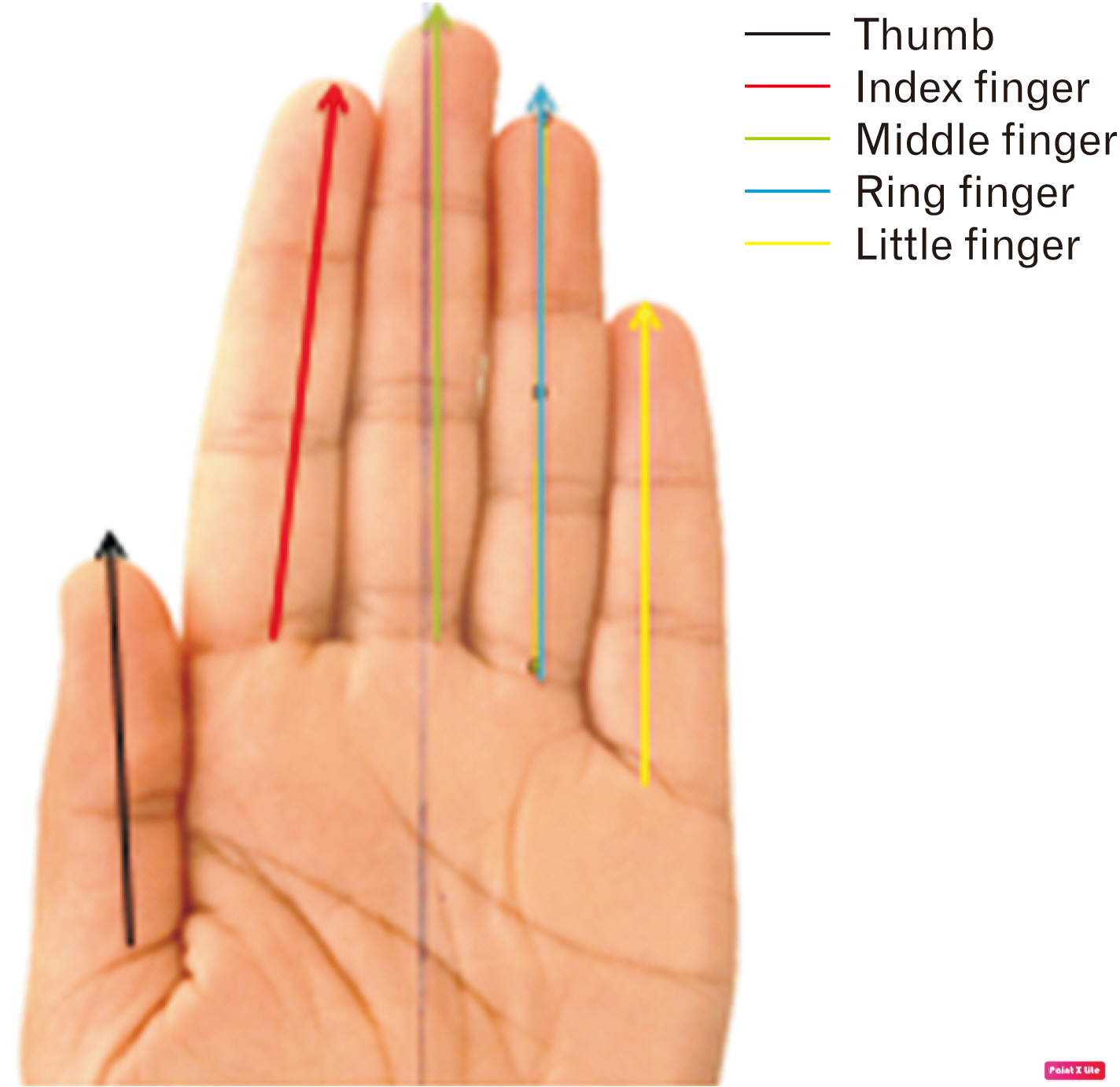
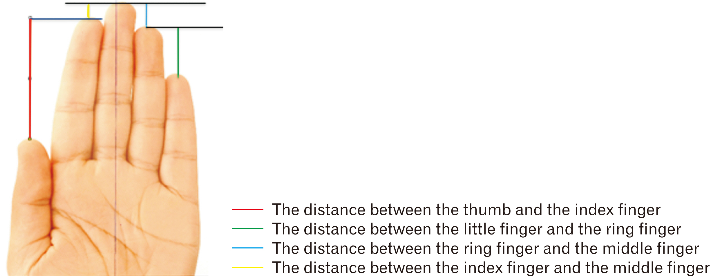
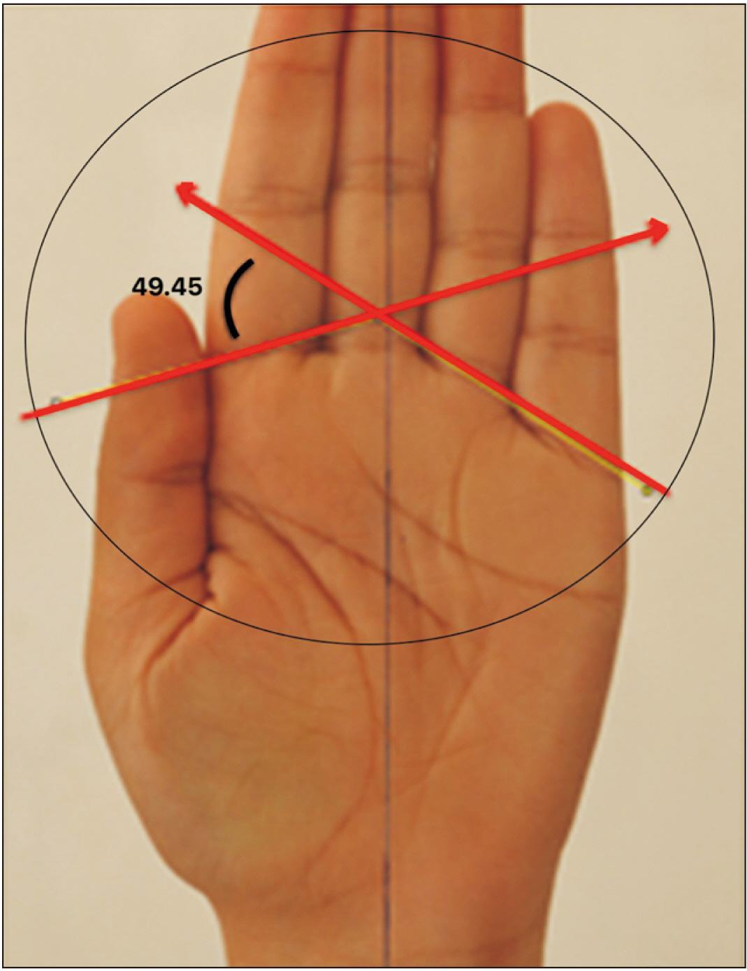
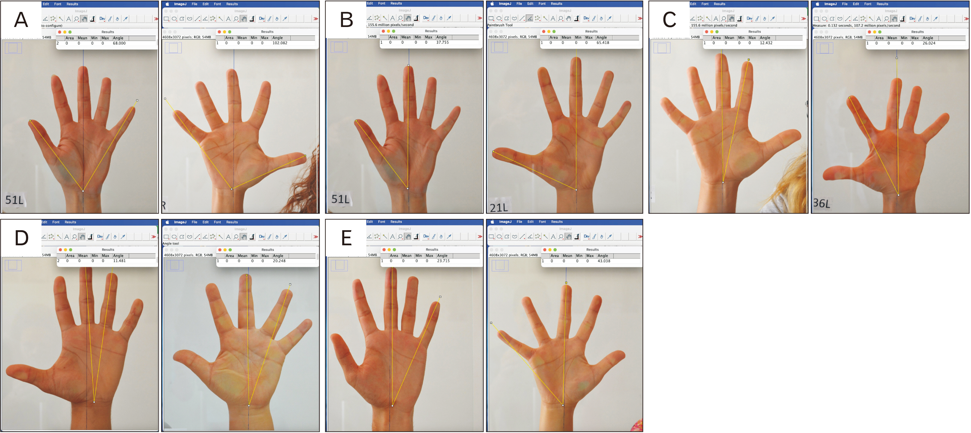
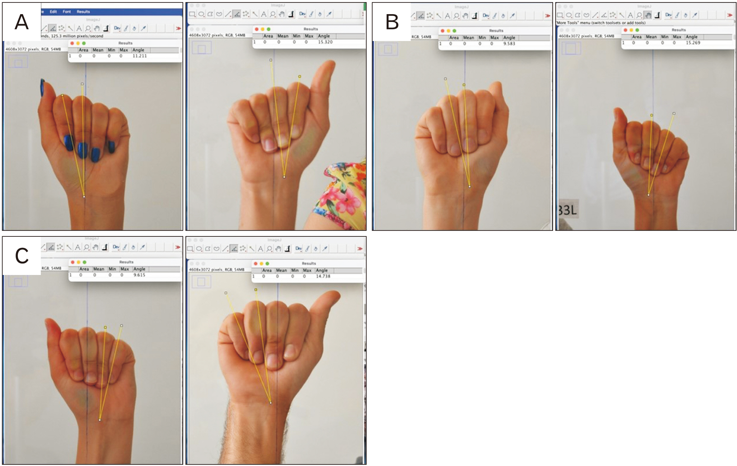
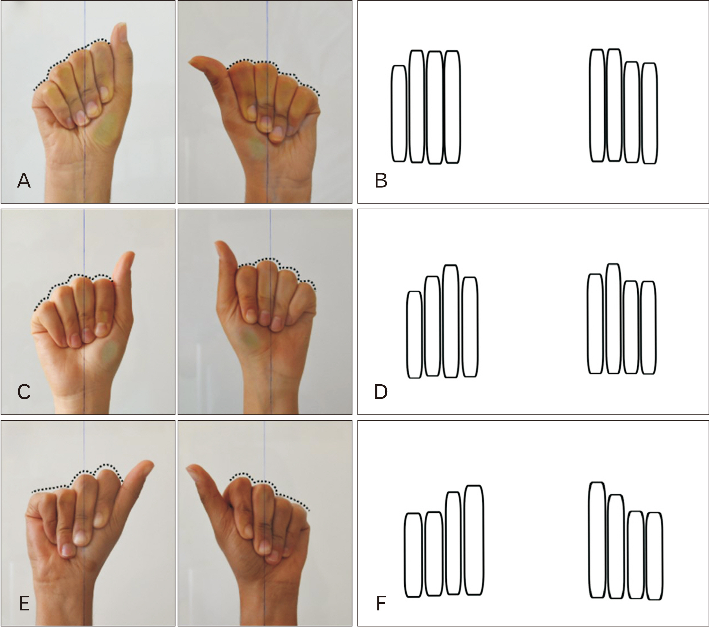
- This study aimed to evaluate the superficial anatomy, kinesiology, and functions of the hand to reveal its morphometry and apply the findings in various fields such as prosthetic hand and protective hand support product design. We examined 51 young adults (32 females, 19 males) aged between 18–30. Hand photographs were taken, and measurements were conducted using ImageJ software. Pearson correlation analysis was performed to determine the relationship between personal information and the parameters. The results of the measurements showed the average lengths of finger segments: thumb (49.5±5.5 mm), index finger (63.9±4.1 mm), middle finger (70.7±5.2 mm), ring finger (65.5±4.8 mm), and little finger (53.3±4.3 mm). Both females and males, the left index finger was measured longer than the right index finger. The right ring finger was found to be longer than the left in both sexes. Additionally, length differences between fingers in extended and maximally adducted positions were determined: thumb-index finger (56.1±6.2 mm), index-middle finger (10.7±4.1 mm), middle-ring finger (10.8±1.4 mm), and ring-little finger (25.6±2.7 mm). Other findings included the average radial natural angle (56.4°±10.5°), ulnar natural angle (23.4°±7.1°), radial deviation angle (65.2°±8.2°), ulnar deviation angle (51.2°±9.6°), and grasping/gripping angle (49.1°±5.8°). The average angles between fingers in maximum abduction positions were also measured: thumb-index finger (53.4°±6.5°), index-middle finger (17.2°±2.6°), middle-ring finger (14.3°±2.3°), and ring-little finger (32.1°±7.0°). The study examined the variability in the positioning of proximal interphalangeal joints during maximum metacarpophalangeal and proximal interphalangeal flexion, coinciding with maximum distal interphalangeal extension movements. The focal points of our observations were the asymmetrical and symmetrical arches formed by these joints. This study provides valuable hand parameters in young adults, which can be utilized in various applications such as prosthetic design, ergonomic product development, and hand-related research. The results highlight the significance of considering individual factors when assessing hand morphology and function.
Keyword
Figure
Reference
-
References
1. Cardoso R, Szabo RM. 2010; Wrist anatomy and surgical approaches. Hand Clin. 26:1–19. DOI: 10.1016/j.hcl.2009.08.009. PMID: 20006240.
Article2. Coelho LA, Gonzalez CLR. 2022; Growing into your hand: the developmental trajectory of the body model. Exp Brain Res. 240:135–45. DOI: 10.1007/s00221-021-06241-2. PMID: 34654947.
Article3. Leversedge FJ. 2008; Anatomy and pathomechanics of the thumb. Hand Clin. 24:219–29. DOI: 10.1016/j.hcl.2008.03.010. PMID: 18675713.
Article4. van Nierop OA, van der Helm A, Overbeeke KJ, Djajadiningrat TJ. 2008; A natural human hand model. Vis Comput. 24:31–44. DOI: 10.1007/s00371-007-0176-x.
Article5. Inouye JM, Valero-Cuevas FJ. 2014; Anthropomorphic tendon-driven robotic hands can exceed human grasping capabilities following optimization. Int J Robotics Res. 33:694–705. DOI: 10.1177/0278364913504247.
Article6. Chatzioglou G, Pinar Y. 2017. The biometric parameters of the hand surface at young adults. Paper presented at: National Anatomy Congress. 2017 Sep 25-27; Abant, Turkey.7. Completo A, Nascimento A, Neto F. 2016; Total arthroplasty of basal thumb joint with Elektra prothesis: an in vitro analysis. J Hand Surg Eur Vol. 41:930–8. DOI: 10.1177/1753193416659230. PMID: 27424207.
Article8. Damert HG, Kober M, Mehling I. 2020; Custom-made wrist prothesis (uni-2™) in a patient with giant cell tumor of the distal radius: 10-year follow-up. Arch Orthop Trauma Surg. 140:2109–14. DOI: 10.1007/s00402-020-03593-2. PMID: 32876750.
Article9. Jones NF, Graham DJ. 2020; Radical resection of a recurrent giant cell tumor of the distal ulna and immediate reconstruction with a distal radio-ulnar joint implant arthroplasty. Hand (N Y). 15:727–31. DOI: 10.1177/1558944719895779. PMID: 31965863. PMCID: PMC7543204.
Article10. Lalchandani GR, Halvorson RT, Zhang AL, Lattanza LL, Immerman I. 2022; Patient outcomes and costs after isolated flexor tendon repairs of the hand. J Hand Ther. 35:590–6. DOI: 10.1016/j.jht.2021.04.015. PMID: 34016517.
Article11. Stival F, Michieletto S, Cognolato M, Pagello E, Müller H, Atzori M. 2019; A quantitative taxonomy of human hand grasps. J Neuroeng Rehabil. 16:28. DOI: 10.1186/s12984-019-0488-x. PMID: 30770759. PMCID: PMC6377750.
Article12. Wanamaker AB, Whelan LR, Farley J, Chaudhari AM. 2019; Biomechanical analysis of users of multi-articulating externally powered prostheses with and without their device. Prosthet Orthot Int. 43:618–28. DOI: 10.1177/0309364619871185. PMID: 31466507.
Article13. Karaçizmeli C, Çakır G, Tükel D. 2014. Robotic hand project. Paper presented at: 2014 22nd Signal Processing and Communications Applications Conference (SIU). 2014 Apr 23-25; Trabzon, Turkey.14. Werner D, Alawi SA. 2019; Four extremity amputation and bionic prosthesis supply after disseminated intravascular coagulation: a follow-up on functionality and quality of life after bionic prosthesis supply. World J Plast Surg. 8:146–62. DOI: 10.29252/wjps.8.2.146. PMID: 31309051. PMCID: PMC6620819.
Article15. Theyskens NC, Vandesande W. Dislocation in single-mobility versus dual-mobility trapezometacarpal joint prostheses. Hand (NY). 2022; Oct. 8. [Epub]. https://doi.org/10.1177/15589447221124257. DOI: 10.1177/15589447221124257. PMID: 36214288.
Article16. Roche AD, Bailey ZK, Gonzalez M, Vu PP, Chestek CA, Gates DH, Kemp SWP, Cederna PS, Ortiz-Catalan M, Aszmann OC. 2023; Upper limb prostheses: bridging the sensory gap. J Hand Surg Eur Vol. 48:182–90. DOI: 10.1177/17531934221131756. PMID: 36649123. PMCID: PMC9996795.
Article17. Demers LAA, Gosselin C. 2011. Kinematic design of a planar and spherical mechanism for the abduction of the fingers of an anthropomorphic robotic hand. Paper presented at: 2011 IEEE International Conference on Robotics and Automation. 2011 May 9-13; Shanghai, China. p. 5350–6.18. Matheson AB, Sinclair DC, Skene WG. 1970; The range and power of ulnar and radial deviation of the fingers. J Anat. 107:439–58.19. Aguilar-Pereyra JF, Castillo-Castaneda E. 2016; Design of a reconfigurable robotic system for flexoextension fitted to hand fingers size. Appl Bionics Biomech. 2016:1712831. DOI: 10.1155/2016/1712831. PMID: 27524880. PMCID: PMC4976261.
Article20. Amundsen A, Rizzo M, Berger RA, Houdek MT, Frihagen F, Moran SL. 2023; Twenty-year experience with primary distal radioulnar joint arthroplasty from a single institution. J Hand Surg Am. 48:53–67. DOI: 10.1016/j.jhsa.2022.02.014. PMID: 35550310.
Article21. Spilman HW, Pinkston D. 1969; Relation of test positions to radial and ulnar deviation. Phys Ther. 49:837–44. DOI: 10.1093/ptj/49.8.837. PMID: 5799806.
Article22. Lee J, Kunii TL. 1995; Model-based analysis of hand posture. IEEE Comp Gr Appl. 15:77–86. DOI: 10.1109/38.403831.
Article23. Billard A, Kragic D. 2019; Trends and challenges in robot manipulation. Science. 364:eaat8414. DOI: 10.1126/science.aat8414. PMID: 31221831.
Article24. Lin J, Wu Y, Huang TS. 2000. Modeling the constraints of human hand motion. Paper presented at: Proceedings Workshop on Human Motion. 2000 Dec 7-8; Austin, TX, USA. p. 121–6.25. Dutagaci H, Sankur B, Yörük E. 2008; Comparative analysis of global hand appearance-based person recognition. J Electron Imaging. 17:011018. DOI: 10.1117/1.2890986.26. Eksioglu M. 2004; Relative optimum grip span as a function of hand anthropometry. Int J Ind Ergon. 34:1–12. DOI: 10.1016/j.ergon.2004.01.007.
Article27. Lippa RA. 2003; Are 2D: 4D finger-length ratios related to sexual orientation? Yes for men, no for women. J Pers Soc Psychol. 85:179–88. DOI: 10.1037/0022-3514.85.1.179. PMID: 12872893.28. Metcalf CD, Notley SV, Chappell PH, Burridge JH, Yule VT. 2008; Validation and application of a computational model for wrist and hand movements using surface markers. IEEE Trans Biomed Eng. 55:1199–210. DOI: 10.1109/TBME.2007.908087. PMID: 18334414.
Article29. Baronio G, Harran S, Signoroni A. 2016; A critical analysis of a hand orthosis reverse engineering and 3D printing process. Appl Bionics Biomech. 2016:8347478. DOI: 10.1155/2016/8347478. PMID: 27594781. PMCID: PMC4993931.
Article30. Hu RX, Jia W, Zhang D, Gui J, Song LT. 2012; Hand shape recognition based on coherent distance shape contexts. Pattern Recognit. 45:3348–59. DOI: 10.1016/j.patcog.2012.02.018.
Article31. Li M, Zhuo Y, He B, Liang Z, Xu G, Xie J, Zhang S. A 3D-printed soft hand exoskeleton with finger abduction assistance. Paper presented at: 2019 16th International Conference on Ubiquitous Robots (UR). 2019 Jun 24-27; Jeju, Korea. p. 319–22. DOI: 10.1109/URAI.2019.8768611.32. Savič T, Pavešić N. 2007; Personal recognition based on an image of the palmar surface of the hand. Pattern Recognit. 40:3152–63. DOI: 10.1016/j.patcog.2007.03.005.
Article33. Park G, Kim S. 2013; Hand biometric recognition based on fused hand geometry and vascular patterns. Sensors (Basel). 13:2895–910. DOI: 10.3390/s130302895. PMID: 23449119. PMCID: PMC3658721.
Article34. Pillai JK, Patel VM, Chellappa R, Ratha NK. 2011; Secure and robust iris recognition using random projections and sparse representations. IEEE Trans Pattern Anal Mach Intell. 33:1877–93. DOI: 10.1109/TPAMI.2011.34. PMID: 21339529.
Article35. Sandow MJ, McMahon M. 2011; Active mobilisation following single cross grasp four-strand flexor tenorrhaphy (Adelaide repair). J Hand Surg Eur Vol. 36:467–75. DOI: 10.1177/1753193411405937. PMID: 21502306.
Article36. Taner HA, Gözil R, İşeri E, Buru E, Bahçelioğlu M. 2016; A comparison of obsessive compulsive disorder, pervasive developmental disorder, and control groups in terms of 2D:4D ratio and finger lengths. GMJ. 27:40–4. Turkish. DOI: 10.1177/1753193411405937.37. Onat P, Kan S, Nakkaş EÇ, Eryılmaz T, Kadayıfçı EC. El boyutlarindan boy tahmininde sosyoekonomik yapinin etkisi. In : XV Student Symposium Working Group Presentations; 2013.38. Ertuğrul B, Otağ İ. 2012; Relationship between sexually dimorphic body morphology and second-to-fourth digit ratio. İnsanbil Derg. 1:94–107. Turkish.
- Full Text Links
- Actions
-
Cited
- CITED
-
- Close
- Share
- Similar articles
-
- Ergonomic Evaluation of Biomechanical Hand Function
- Myoelectric Hand Rehabilitation in the Bilateral Upper Extremity Amputee of Burn Patients: Two Cases Report
- A Case Report of Living Transplantation for Post-traumatic Ankylosis of 2nd M-P Joint of the Hand
- A clinical analysis on hand injuries
- Merger of Two Journals


