Arch Hand Microsurg.
2024 Jun;29(2):116-121. 10.12790/ahm.23.0058.
Surgical treatment of extensive and multiple skin cancers via excision and reconstruction using multiple flaps: a case report
- Affiliations
-
- 1Department of Plastic Surgery, Samsung Medical Centre, Sungkyunkwan University School of Medicine, Seoul, Korea
- KMID: 2556362
- DOI: http://doi.org/10.12790/ahm.23.0058
Abstract
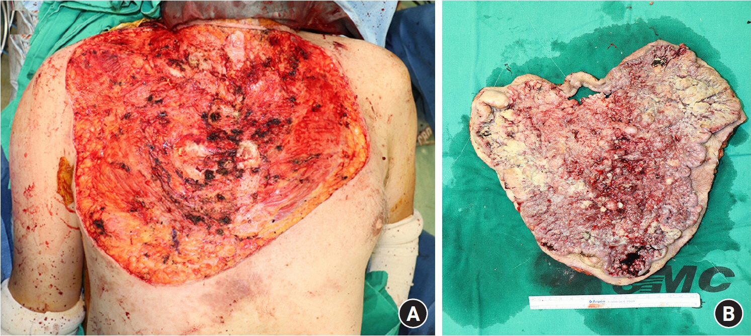
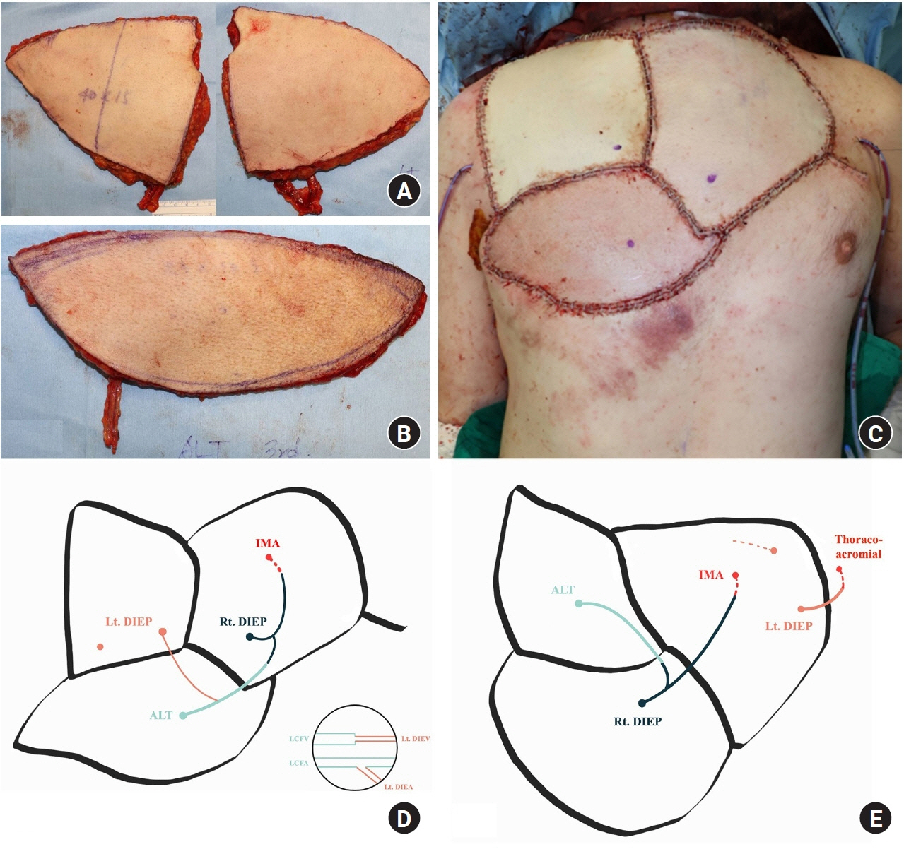
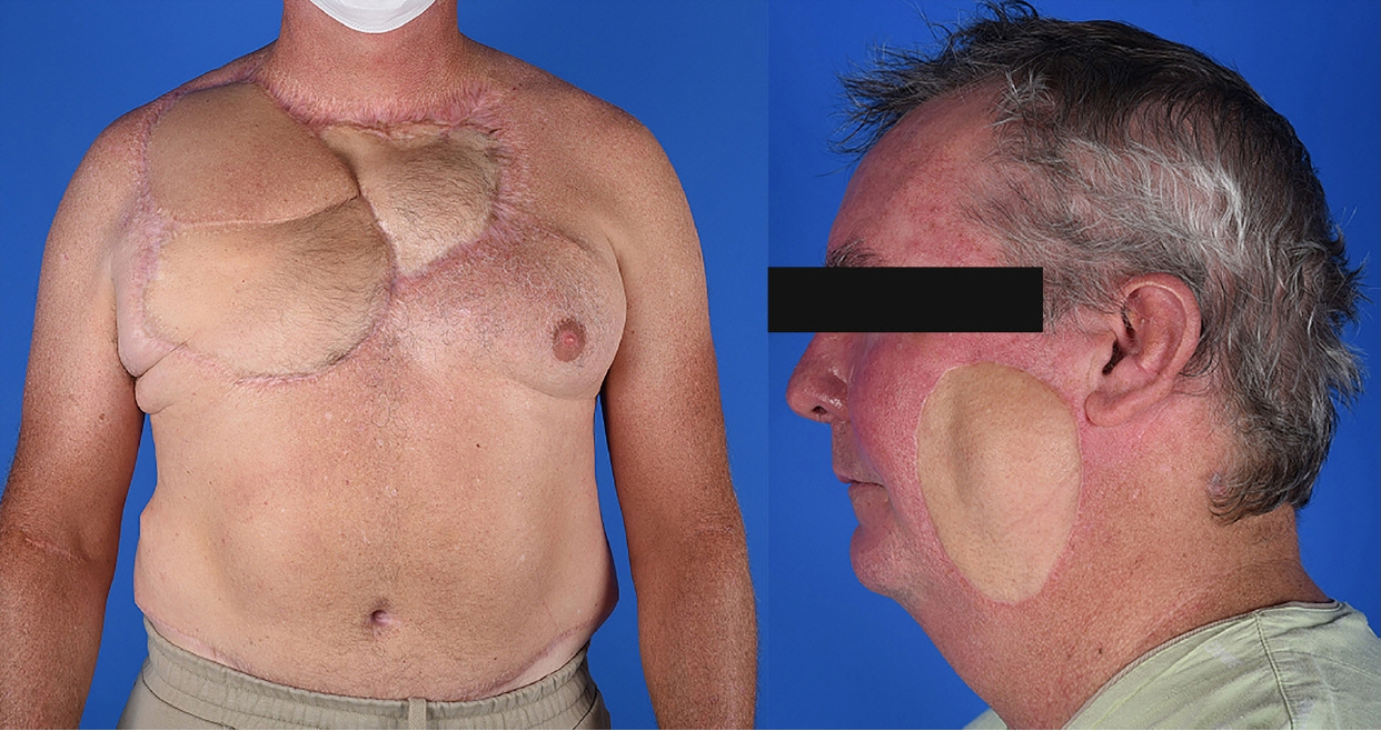
- A 47-year-old male patient presented with multiple squamous and basal cell carcinomas on the anterior chest, back, and left cheek. The patient experienced odorous discharge from the tumors. Surgical excision was planned, beginning with the anterior chest squamous cell carcinoma. An extensive 32×30 cm cutaneous defect was created, which was covered by a bilateral deep inferior epigastric perforator and pedicled latissimus dorsi myocutaneous flaps. The basal cell carcinomas on the back and squamous cell carcinoma on the left cheek were serially excised, after which the left cheek wound required flap coverage. Postoperative complications such as venous thrombosis and infection led to several reoperations, yet the extensive defect was successfully reconstructed. No local recurrence developed during 31 months of follow-up. We report this case to demonstrate that although the wide excision of very large skin cancers may result in extensive and challenging defects as large as 5.8% of the total body surface area, coverage with appropriate flaps may lead to successful oncologic outcomes and improve the patient’s quality of life.
Figure
Reference
-
References
1. Siegel RL, Miller KD, Wagle NS, Jemal A. Cancer statistics, 2023. CA Cancer J Clin. 2023; 73:17–48.
Article2. Motley R, Kersey P, Lawrence C; British Association of Dermatologists; British Association of Plastic Surgeons. Multiprofessional guidelines for the management of the patient with primary cutaneous squamous cell carcinoma. Br J Plast Surg. 2003; 56:85–91.
Article3. Jarkowski A 3rd, Hare R, Loud P, et al. Systemic therapy in advanced cutaneous squamous cell carcinoma (CSCC): the Roswell Park experience and a review of the literature. Am J Clin Oncol. 2016; 39:545–8.4. Bird C. Managing malignant fungating wounds. Prof Nurse. 2000; 15:253–6.5. Pyo S, Joo H. 479 Vaccum-assisted closure therapy in split-thickness skin graft on the wound on the contours of the body. J Burn Care Res. 2019; 40(Suppl 1):S214–5.
Article6. Grocott P, Gethin G, Probst S. Malignant wound management in advanced illness: new insights. Curr Opin Support Palliat Care. 2013; 7:101–5.7. Sim NHS, Ngaserin S, Feng JJ, et al. Reconstruction of a massive chest wall defect using a free anterolateral-lower medial thigh flaps: a case report. J Surg Case Rep. 2023; 2023:rjad264.
Article
- Full Text Links
- Actions
-
Cited
- CITED
-
- Close
- Share
- Similar articles
-
- Management of Facial Skin Cancer: Surgical Excision and Immediate Reconstruction
- Reconstruction of Large Facial Defects via Excision of Skin Cancer Using Two or More Regional Flaps
- Reconstructive Modalities According to Aesthetic Consideration of Subunits of the Cheek after Wide Excision of Skin Cancer
- Chimeric anterolateral thigh flap for reconstruction of complex defects in oral cancer: a report of three cases
- A clinical review of reconstructive techniques for patients with multiple skin cancers on the face


