Arch Hand Microsurg.
2024 Jun;29(2):105-109. 10.12790/ahm.24.0006.
Neglected very large ancient schwannoma of the distal wrist: a case report and literature review
- Affiliations
-
- 1Department of Plastic and Reconstructive Surgery, Saeson Hospital, Daejeon, Korea
- 2Department of Orthopaedic Surgery, Saeson Hospital, Daejeon, Korea
- 3Research Institute of Clinical Medicine, Woori Madi Medical Center, Jeonju, Korea
- KMID: 2556360
- DOI: http://doi.org/10.12790/ahm.24.0006
Abstract
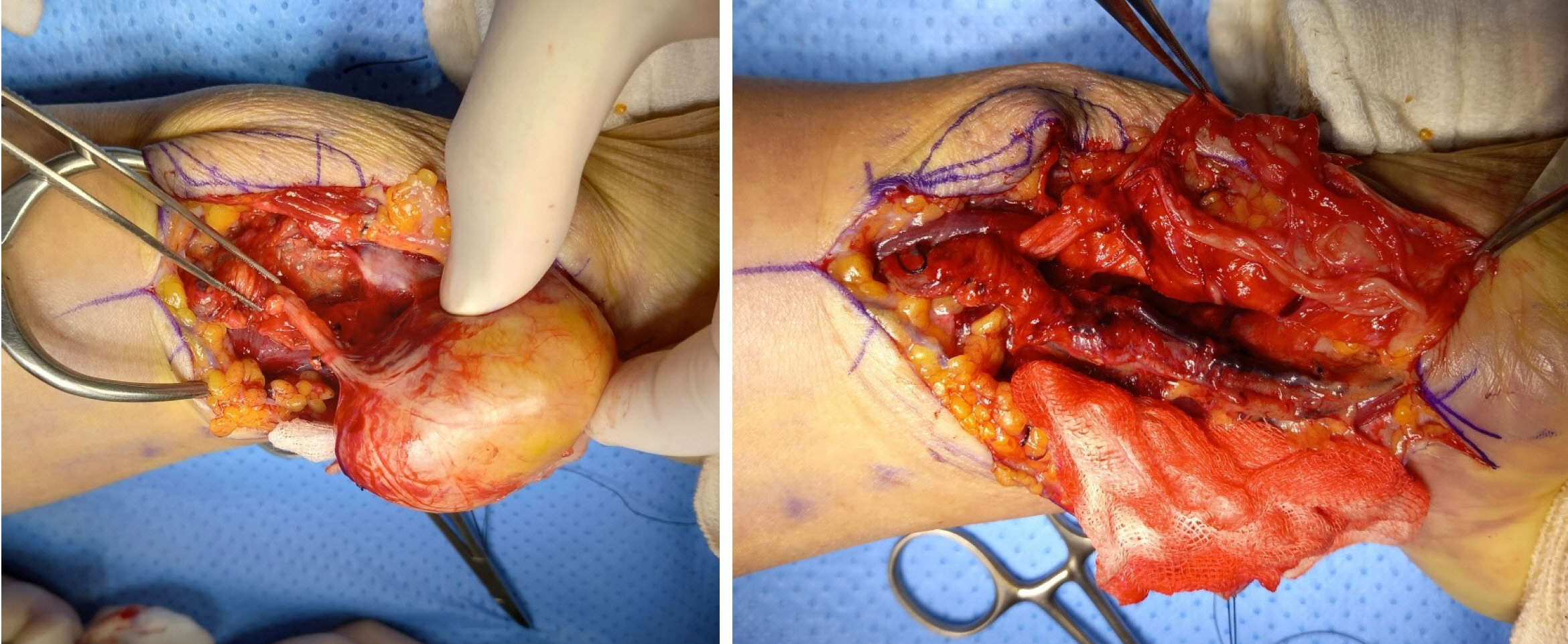
- Ancient schwannoma is a variant of schwannoma characterized by slow progression, degenerative changes, and a higher incidence in older adults. There have been two prior reported cases of ancient schwannoma arising from the distal ulnar nerve at the wrist level, but neither were longstanding or very large. Herein, we report an ancient schwannoma found in the ulnar nerve of the distal forearm that was found to be clinically meaningful in size. A 61-year-old man presented with complaints of tingling sensation of the fourth and fifth fingers and bulging of the ulnar side of the wrist. The patient reported that the mass in his wrist had grown very slowly, starting about 10 years ago, and that he had started experiencing a tingling sensation in his fourth and fifth fingers about 3 years prior, which had become worse in the past year. Based on the results of the preoperative examination, a benign nerve sheath tumor was suspected. As it was thought that the possibility of malignancy was not high, we elected to perform a marginal excision. Pathological examination confirmed ancient schwannoma. At his most recent visit, 3 years after surgery, he reported no recurrence and that he felt better than before surgery, but some tingling sensations remained. As with small ancient schwannoma in the distal wrist, most cases of large ancient schwannoma can be treated without special complications based on an accurate preoperative diagnosis.
Keyword
Figure
Reference
-
References
1. Ackerman LV, Taylor FH. Neurogenous tumors within the thorax; a clinicopathological evaluation of forty-eight cases. Cancer. 1951; 4:669–91.
Article2. Joseph CM, Cherian M, Mishra MN, Chase A. Ancient schwannoma of the distal ulnar nerve: a rare presentation. J Clin Mol Pathol. 2020; 4:24.3. Yoon T, Hong KY. Multiple ancient schwannomas of the ulnar nerve at distant sites: a case report. Arch Hand Microsurg. 2022; 27:88–92.
Article4. Kehoe NJ, Reid RP, Semple JC. Solitary benign peripheral-nerve tumours. Review of 32 years' experience. J Bone Joint Surg Br. 1995; 77:497–500.
Article5. Ogose A, Hotta T, Morita T, et al. Tumors of peripheral nerves: correlation of symptoms, clinical signs, imaging features, and histologic diagnosis. Skeletal Radiol. 1999; 28:183–8.
Article6. Dodd LG, Marom EM, Dash RC, Matthews MR, McLendon RE. Fine-needle aspiration cytology of "ancient" schwannoma. Diagn Cytopathol. 1999; 20:307–11.
Article7. Bhattacharyya AK, Perrin R, Guha A. Peripheral nerve tumors: management strategies and molecular insights. J Neurooncol. 2004; 69:335–49.
Article8. Lee YS, Kim JO, Park SE. Ancient schwannoma of the thigh mimicking a malignant tumour: a report of two cases, with emphasis on MRI findings. Br J Radiol. 2010; 83:e154–7.
Article9. Sawada T, Sano M, Ogihara H, Omura T, Miura K, Nagano A. The relationship between pre-operative symptoms, operative findings and postoperative complications in schwannomas. J Hand Surg Br. 2006; 31:629–34.
Article
- Full Text Links
- Actions
-
Cited
- CITED
-
- Close
- Share
- Similar articles
-
- A Case of Ancient Schwannoma of the Lingual Nerve
- Ancient schwannoma in the parotid gland: A case report and review of the literature
- Multiple ancient schwannomas of the ulnar nerve at distant sites: a case report
- A Case of Ancient Schwannoma of the Submandibular Gland
- Solitary Ancient Schwannoma in Upper Arm: A Case Report


