Arch Hand Microsurg.
2024 Jun;29(2):90-95. 10.12790/ahm.24.0014.
The Zitelli bilobed flap for soft tissue coverage after mucoid cyst resection: a retrospective cohort study
- Affiliations
-
- 1Department of Plastic and Reconstructive Surgery, Saeson Hospital, Daejeon, Korea
- 2Department of Plastic and Reconstructive Surgery, Keimyung University Dongsan Hospital, Daegu, Korea
- KMID: 2556358
- DOI: http://doi.org/10.12790/ahm.24.0014
Abstract
- Purpose
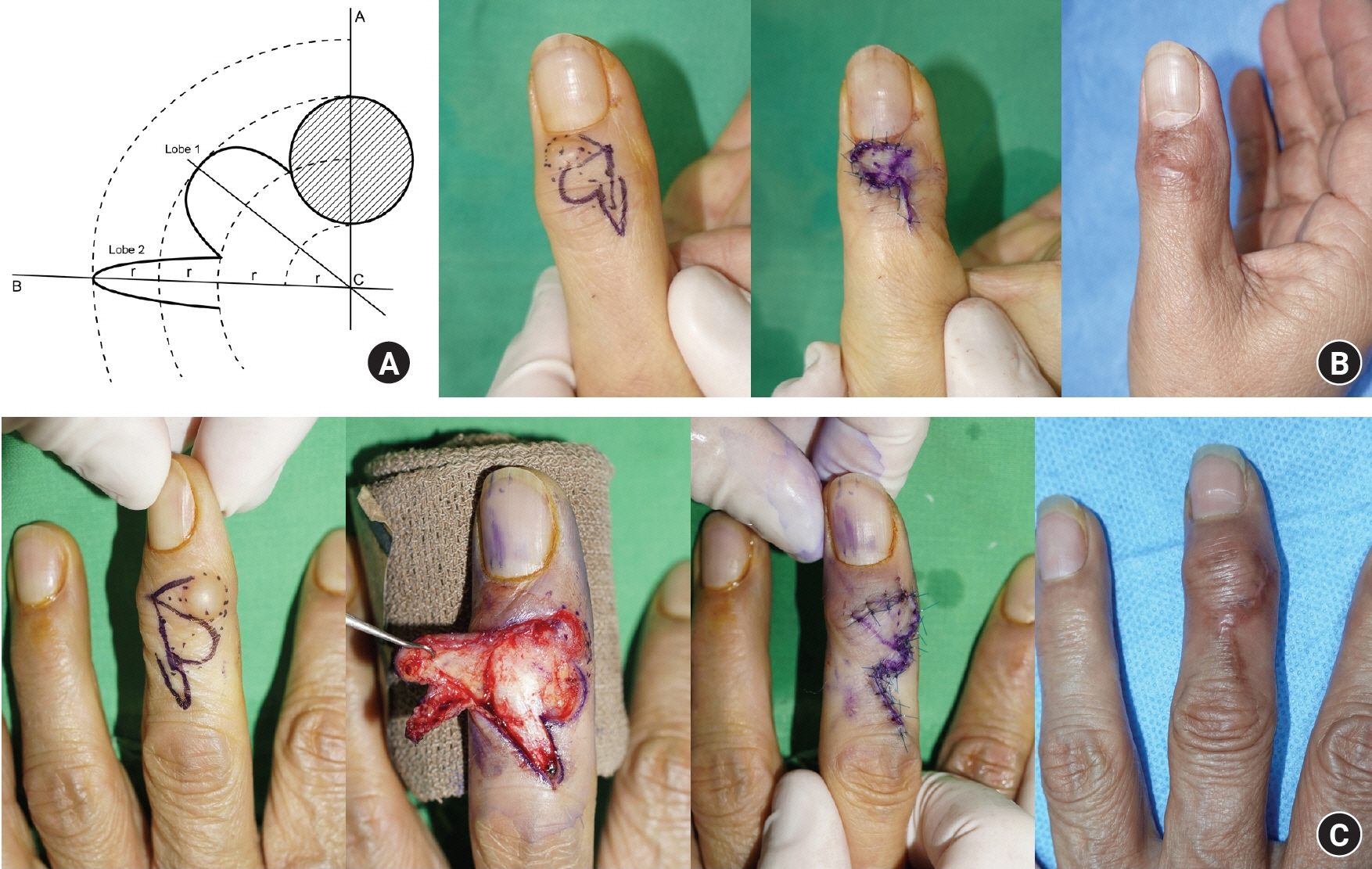
Digital mucoid cysts are frequently found at the distal interphalangeal (DIP) joint in patients with degenerative osteoarthritis. In complicated cases, surgical treatment with mucoid cyst resection is considered, and soft tissue is covered with one of various local flap techniques. Among these, bilobed flaps are reliable and aesthetically favorable, with primary healing of the donor site. In this study, we investigated a case series of bilobed flaps for digital mucoid cysts.
Methods
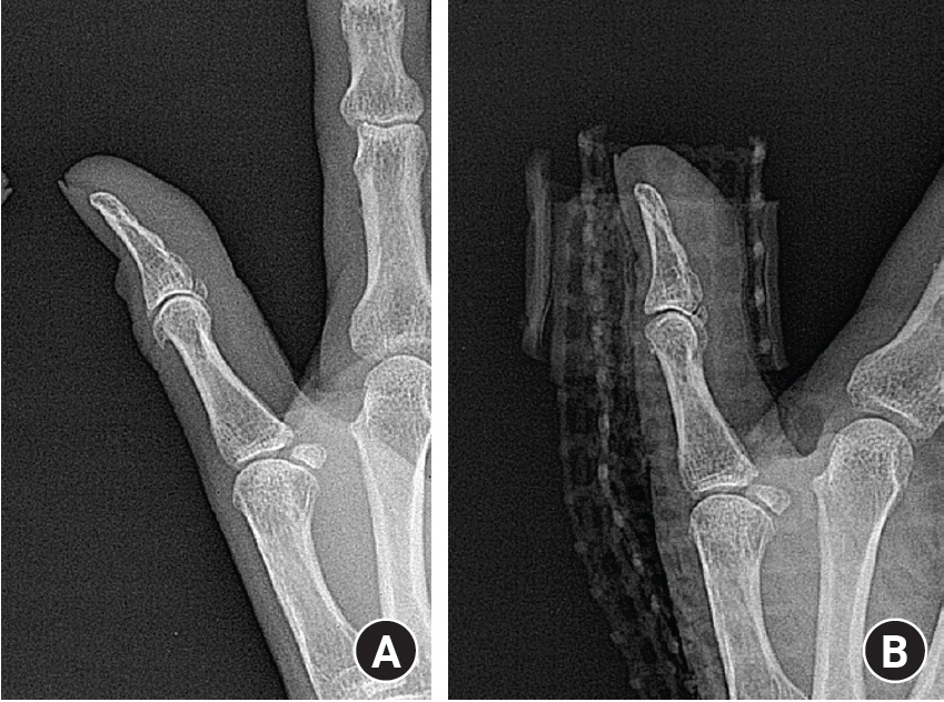
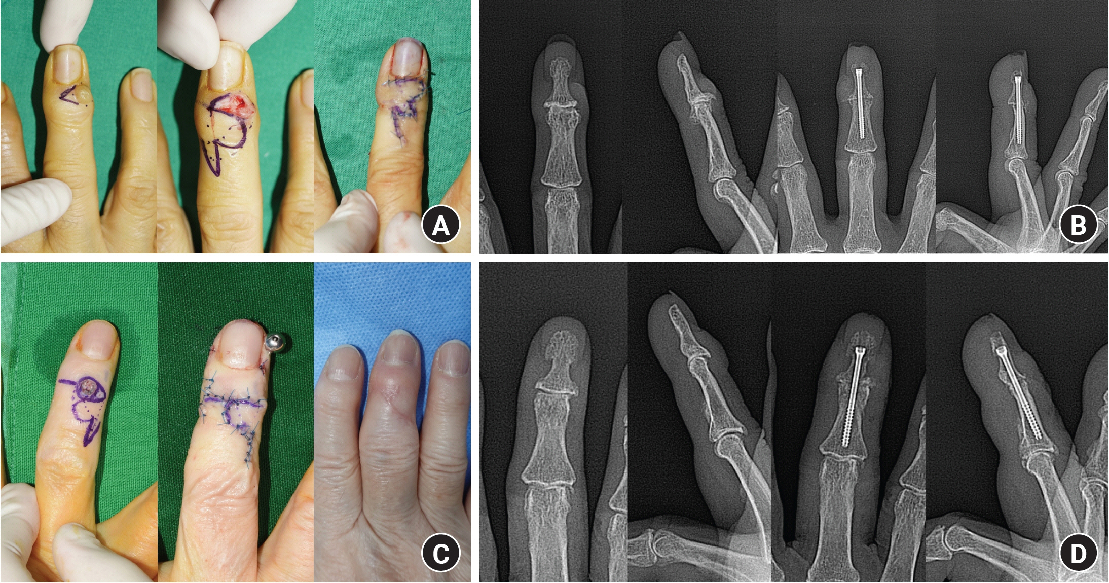
We retrospectively reviewed our electronic medical records and found 26 digital mucoid cysts treated with bilobed flaps at our facility between July 2022 and February 2024. We extracted data from the records of these patients on sex; age; time to surgery; clinical findings including nail ridging, the presence of osteophytes, cyst size and location, and additional procedures (arthrodesis); and follow-up data including the occurrence of complications, such as delayed wound healing, infection, stiffness, and recurrence.
Results
Among the 26 patients in our sample, 19 were female and seven were male. The average age was 62.2 years, and the average time to surgery was 10.8 months. Preoperatively, the average cyst measured 6.9×8.3 mm. Nail ridging was found in 19 patients (73.1%) and osteophytes in 22 patients (84.6%). The most commonly affected digit was the middle finger, which accounted for 10 cases (38.5%). All the flaps totally survived, without major complications.
Conclusion
Based on our series, a bilobed flap for soft tissue coverage after mucoid cyst excision can achieve high-quality surgical results.
Keyword
Figure
Reference
-
References
1. Rizzo M, Beckenbaugh RD. Treatment of mucous cysts of the fingers: review of 134 cases with minimum 2-year follow-up evaluation. J Hand Surg Am. 2003; 28:519–24.
Article2. Young KA, Campbell AC. The bilobed flap in treatment of mucous cysts of the distal interphalangeal joint. J Hand Surg Br. 1999; 24:238–40.
Article3. Blume PA, Moore JC, Novicki DC. Digital mucoid cyst excision by using the bilobed flap technique and arthroplastic resection. J Foot Ankle Surg. 2005; 44:44–8.
Article4. Jiménez I, Delgado PJ, Kaempf de Oliveira R. The Zitelli bilobed flap on skin coverage after mucous cyst excision: a retrospective cohort of 33 cases. J Hand Surg Am. 2017; 42:506–10.
Article5. Kim EJ, Huh JW, Park HJ. Digital mucous cyst: a clinical-surgical study. Ann Dermatol. 2017; 29:69–73.
Article6. Li K, Barankin B. Digital mucous cysts. J Cutan Med Surg. 2010; 14:199–206.
Article7. Fritz GR, Stern PJ, Dickey M. Complications following mucous cyst excision. J Hand Surg Br. 1997; 22:222–5.
Article8. Kasdan ML, Stallings SP, Leis VM, Wolens D. Outcome of surgically treated mucous cysts of the hand. J Hand Surg Am. 1994; 19:504–7.
Article9. Dodge LD, Brown RL, Niebauer JJ, McCarroll HR Jr. The treatment of mucous cysts: long-term follow-up in sixty-two cases. J Hand Surg Am. 1984; 9:901–4.
Article10. Zimany A. The bi-lobed flap. Plast Reconstr Surg (1946). 1953; 11:424–34.
Article11. Kleinert HE, Kutz JE, Fishman JH, McCraw LH. Etiology and treatment of the so-called mucous cyst of the finger. J Bone Joint Surg Am. 1972; 54:1455–8.
Article
- Full Text Links
- Actions
-
Cited
- CITED
-
- Close
- Share
- Similar articles
-
- Inguinal Soft Tissue Reconstruction Using Pedicled Anterolateral Thigh Flap: A Case Report
- Upside-down Adipofascial Flap for the Medial Foot Soft Tissue Defect after Trauma: Case Report
- Soft Tissue Coverage Using a Combined Gastrocnemius-medial Sural Artery Perforator Flap
- The Bilobed Flap for Nasal Reconstruction
- Reconstruction of Scalp Defects using Bilobed Flap


