Ewha Med J.
2024 Apr;47(2):e25. 10.12771/emj.2024.e25.
The use of the bicipital groove as an intraoperative landmark for proximal humeral rotation during fracture fixation
- Affiliations
-
- 1Department of Orthopaedic Surgery, School of Medicine, Kyungpook National University, Kyungpook National University Hospital, Daegu, Korea
- 2AIRS, Daegu, Korea
- 3Department of Orthopedic Surgery, St. Carolus Hospital, Faculty of Medicine, Universitas Trisakti, Jakarta, Indonesia
- 4Department of Orthopaedic Surgery, Gyeongsang National University Changwon Hospital, Changwon, Korea
- 5Department of Hand Surgery, Affiliated Hospital of Nantong University, Nantong, China
- 6Department of Orthopaedic Surgery, Asan Medical Center, School of Medicine, University of Ulsan, Seoul, Korea
- KMID: 2556314
- DOI: http://doi.org/10.12771/emj.2024.e25
Abstract
Objectives
This study aimed to quantify the relationship between proximal humeral rotation and the lateral border of the bicipital groove on fluoroscopic imaging.
Methods
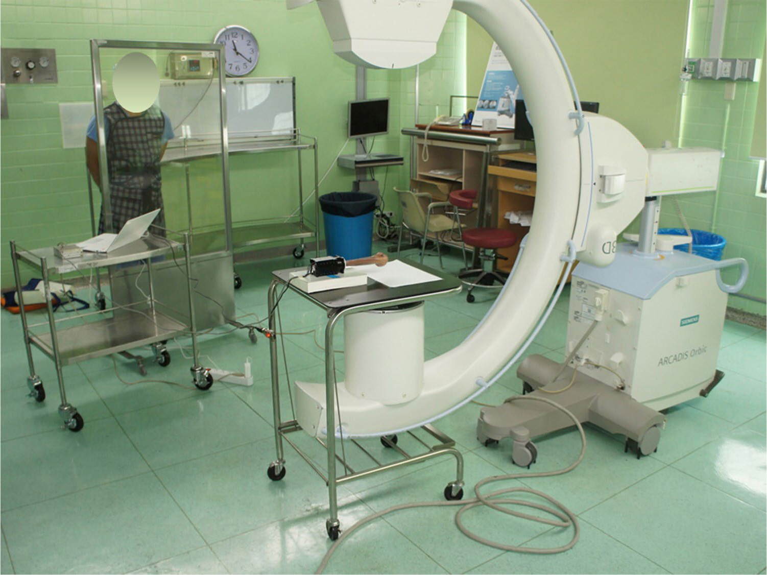
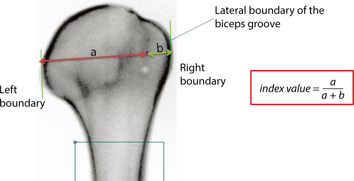
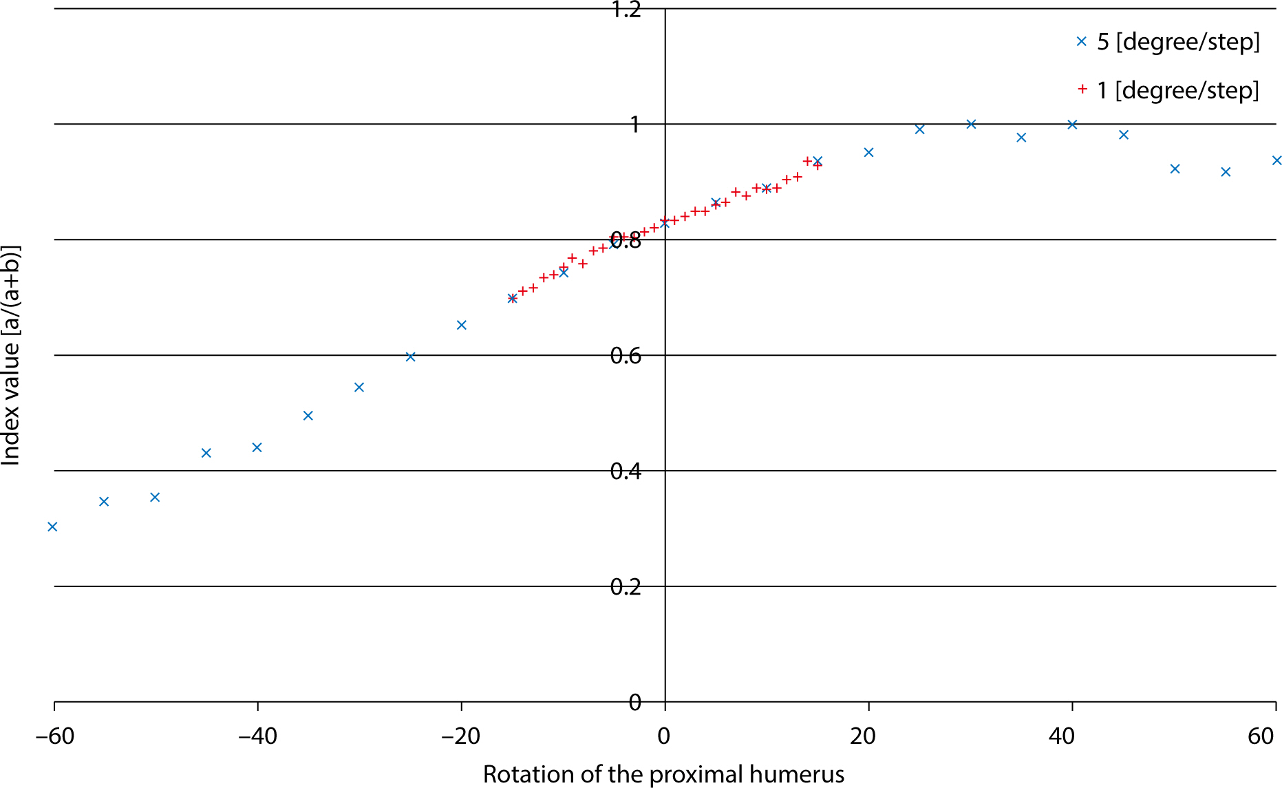
A composite normal humerus with a marker placed on the lateral border of the bicipital groove was affixed to a custom rotation device at the proximal cut segment. Consecutive fluoroscopic images were captured from −60° to 60° in 5° increments and from −15° to 15° in 1° increments. The index value was calculated by taking the ratio of the distance from the medial boundary of the proximal humerus to the lateral border of the bicipital groove to the distance between the medial and lateral boundaries of the proximal humerus. The correlation between the humeral rotation and the index value was determined.
Results
The index value showed a strong positive linear correlation position during internal rotation of the humerus across the entire range (r=0.998, P<0.001), as well as when the humerus was externally rotated, ranging from 15° of internal rotation to 15° of external rotation (r=0.991, P<0.001).
Conclusion
The lateral border of the bicipital groove may serve as a useful intraoperative landmark for assessing proximal humeral rotation. This could potentially enhance the outcomes of humeral fracture repair and upper arm arthroplasty.
Keyword
Figure
Reference
-
References
1. Zhang Q, Liu H, Chen W, Li X, Song Z, Pan J, et al. Radiologic measurement of lesser trochanter and its clinical significance in Chinese. Skelet Radiol. 2009; 38(12):1175–1181. DOI: 10.1007/s00256-009-0662-5. PMID: 19277649.2. Kim JJ, Kim E, Kim KY. Predicting the rotationally neutral state of the femur by comparing the shape of the contralateral lesser trochanter. Orthopedics. 2001; 24(11):1069–1070. DOI: 10.3928/0147-7447-20011101-18. PMID: 11727805.3. Tan J, Lee HJ, Aminata I, Chun JM, Kekatpure AL, Jeon IH. Radiographic landmark for humeral head rotation: a new radiographic landmark for humeral fracture fixation. Injury. 2015; 46(4):666–670. DOI: 10.1016/j.injury.2014.10.059. PMID: 25467709.4. Li Y, Wang C, Wang M, Huang L, Huang Q. Postoperative malrotation of humeral shaft fracture after plating compared with intramedullary nailing. J Shoulder Elbow Surg. 2011; 20(6):947–954. DOI: 10.1016/j.jse.2010.12.016. PMID: 21440461.5. Park SJ, Kim E, Jeong HJ, Lee J, Park S. Prediction of the rotational state of the humerus by comparing the contour of the contralateral bicipital groove: method for intraoperative evaluation. Indian J Orthop. 2012; 46(6):675–679. DOI: 10.4103/0019-5413.104210. PMID: 23325971. PMCID: PMC3543886.6. Wang C, Li J, Li Y, Dai G, Wang M. Is minimally invasive plating osteosynthesis for humeral shaft fracture advantageous compared with the conventional open technique? J Shoulder Elbow Surg. 2015; 24(11):1741–1748. DOI: 10.1016/j.jse.2015.07.032. PMID: 26480879.7. Rommens PM, Blum J, Runkel M. Retrograde nailing of humeral shaft fractures. Clin Orthop Relat Res. 1998; 350:26–39. DOI: 10.1097/00003086-199805000-00004.8. Itoi E, King GJW, Niebur GL, Morrey BF, An KN. Malrotation of the humeral component of the capitellocondylar total elbow replacement is not the sole cause of dislocation. J Orthop Res. 1994; 12(5):665–671. DOI: 10.1002/jor.1100120509. PMID: 7931783.9. Sabo MT, Athwal GS, King GJW. Landmarks for rotational alignment of the humeral component during elbow arthroplasty. J Bone Joint Surg. 2012; 94(19):1794–1800. DOI: 10.2106/JBJS.J.01740. PMID: 23032590.10. Lee HJ, Oh CW, Oh JK, Apivatthakakul T, Kim JW, Yoon JP, et al. Minimally invasive plate osteosynthesis for humeral shaft fracture: a reproducible technique with the assistance of an external fixator. Arch Orthop Trauma Surg. 2013; 133(5):649–657. DOI: 10.1007/s00402-013-1708-7. PMID: 23463256.11. Boileau P, Bicknell RT, Mazzoleni N, Walch G, Urien JP. CT scan method accurately assesses humeral head retroversion. Clin Orthop Relat Res. 2008; 466(3):661–669. DOI: 10.1007/s11999-007-0089-z. PMID: 18264854. PMCID: PMC2505224.12. Vlachopoulos L, Carrillo F, Dunner C, Gerber C, Székely C, Fürnstahl P. A novel method for the approximation of humeral head retrotorsion based on three-dimensional registration of the bicipital groove. J Bone Joint Surg. 2018; 100(15):e101. DOI: 10.2106/JBJS.17.01561. PMID: 30063597.13. Micic I, Kholinne E, Hong H, Choi H, Kwak JM, Sun Y, et al. Navigation-assisted suture anchor insertion for arthroscopic rotator cuff repair. BMC Musculoskelet Disord. 2019; 20(1):633. DOI: 10.1186/s12891-019-3021-2. PMID: 31884952. PMCID: PMC6935480.
- Full Text Links
- Actions
-
Cited
- CITED
-
- Close
- Share
- Similar articles
-
- Measurement of Proximal Humerus in Korean Adult Skeleton
- Conformity of Proximal Humerus Internal Locking Plate System (PHILOS) and Anatomic Features of Proximal Locking Screws: Cadaveric Study
- Analysis of Anatomical Conformity of Straight Antegrade Humeral Intramedullary Nail in Korean
- Biomechanical investigation of arm position on deforming muscular forces in proximal humerus fractures
- Medial and Lateral Dual Plate Fixation for Osteoporotic Proximal Humerus Comminuted Fracture: 2 Case Reports


