Early diagnosis of negative-pressure pulmonary edema presenting as diffuse alveolar hemorrhage using lung ultrasonography -A case report-
- Affiliations
-
- 1Department of~, Ulsan University Hospital, University of Ulsan College of Medicine, Ulsan, Korea
- KMID: 2555251
- DOI: http://doi.org/10.17085/apm.23101
Abstract
- Background
Diffuse alveolar hemorrhage (DAH) is a potentially life-threatening condition that can occur due to a variety of disorders. Hence, rapid diagnosis and prompt initiation of appropriate treatment are imperative. Case: A 55-year-old woman with a deep neck infection underwent emergent tonsillectomy. General anesthesia and surgery proceeded uneventfully. Upon transfer to the post-anesthesia care unit, ongoing respiratory distress and occasional expectoration of blood-tinged sputum were noted. Lung ultrasonography (LUS) revealed multiple B-profiles and irregular pleural lines with subpleural consolidations. Emergent bronchoscopy with bronchoalveolar lavage was diagnostic of DAH. She underwent a comprehensive evaluation for rheumatologic and infectious etiologies of DAH, all of which yielded negative results. The patient was managed with steroids and conservative treatment.
Conclusions
The integration of LUS with clinical information allows for more rapid differentiation of acute respiratory failure causes. Therefore, anesthesiologists’ awareness and utilization of LUS findings of DAH can significantly contribute to appropriate management.
Keyword
Figure
-
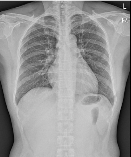
Fig. 1. Chest X-ray depicted no pathological findings–despite the patient’s symptoms.
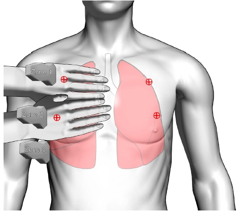
Fig. 2. The procedure of lung ultrasonography. The patient was examined in the supine position, with the probe being placed on the bedside lung ultrasound in emergency point, especially on the anterior chest, to confirm the ultrasound findings.
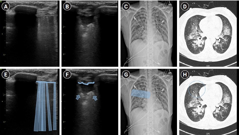
Fig. 3. An integrated assessment of Type 2 negative-pressure pulmonary edema, presenting as diffuse alveolar hemorrhage, using lung ultrasound (A, B), chest radiograph (C), and high–resolution computed tomography (HRCT) (D) with schematic representations (E–H). The infiltrative opacification pattern observed in the mid-zone on the chest radiograph (C) and the extensive ground-glass opacities and consolidations seen on the HRCT (D) corresponds to multiple B-lines (A, E) and irregular pleural line with subpleural consolidation (B, F) detected on lung ultrasound. The detailed examination of the lung ultrasound scan is represented by the blue box (G) and blue circle (H), with the convex probe (A, E) and linear probe (B, F) placed longitudinally.
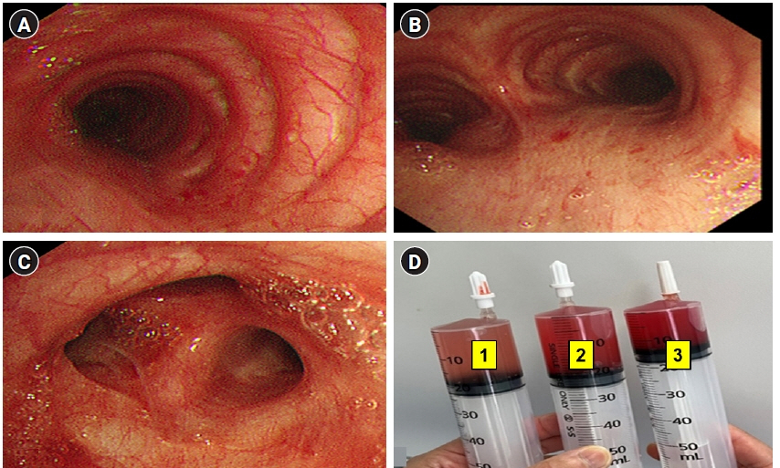
Fig. 4. (A) Diffuse alveolar hemorrhage (DAH) in bronchoalveolar lavage (BAL), at trachea (B) DAH in BAL, at carina (C) DAH in BAL at the lingual region of the left upper lobe (D) The progressive hemorrhagic BAL in the serial samples (number 1 is the first BAL, number 2 is the second BAL, and number 3 is the third BAL).
Reference
-
1. Tamai K, Tomii K, Nakagawa A, Otsuka K, Nagata K. Diffuse alveolar hemorrhage with predominantly right-sided infiltration resulting from cardiac comorbidities. Intern Med. 2015; 54:319–24.2. Lara AR, Schwarz MI. Diffuse alveolar hemorrhage. Chest. 2010; 137:1164–71.3. Robbins RA, Linder J, Stahl MG, Thompson AB, Haire W, Kessinger A, et al. Diffuse alveolar hemorrhage in autologous bone marrow transplant recipients. Am J Med. 1989; 87:511–8.4. Zhou T, Zhang HP, Chen WW, Xiong ZY, Fan T, Fu JJ, et al. Cuff-leak test for predicting postextubation airway complications: a systematic review. J Evid Based Med. 2011; 4:242–54.5. Lichtenstein DA. BLUE-protocol and FALLS-protocol: two applications of lung ultrasound in the critically ill. Chest. 2015; 147:1659–70.6. De Prost N, Parrot A, Cuquemelle E, Picard C, Antoine M, Fleury-Feith J, et al. Diffuse alveolar hemorrhage in immunocompetent patients: etiologies and prognosis revisited. Respir Med. 2012; 106:1021–32.
Article7. Alexandre AT, Vale A, Gomes T. Diffuse alveolar hemorrhage: how relevant is etiology? Sarcoidosis Vasc Diffuse Lung Dis. 2019; 36:47–52.8. Buda N, Masiak A, Smoleńska Ż, Gałecka K, Porzezińska M, Zdrojewski Z. Serial lung ultrasonography to monitor patient with diffuse alveolar hemorrhage. Ultrasound Q. 2017; 33:86–9.9. Austin A, Modi A, Judson MA, Chopra A. Sevoflurane induced diffuse alveolar hemorrhage in a young patient. Respir Med Case Rep. 2016; 20:14–5.
Article10. Choe YS, Kim NY, Lee AR. Diffuse alveolar hemorrhage in patients undergoing neurointervention: a case report. Anesth Pain Med. 2016; 6:e33979.
Article11. Kim JP, Park JJ, Kim NJ, Woo SH. A case of diffuse alveolar hemorrhage after tonsillectomy -a case report-. Korean J Anesthesiol. 2012; 63:165–8.
Article12. Bhaskar B, Fraser JF. Negative pressure pulmonary edema revisited: pathophysiology and review of management. Saudi J Anaesth. 2011; 5:308–13.13. Schwartz DR, Maroo A, Malhotra A, Kesselman H. Negative pressure pulmonary hemorrhage. Chest. 1999; 115:1194–7.
Article14. Kim CA, Liu R, Hsia DW. Diffuse alveolar hemorrhage induced by sevoflurane. Ann Am Thorac Soc. 2014; 11:853–5.
Article
- Full Text Links
-
- Actions
-
Cited
- CITED
-
- Close
- Share
-
- Similar articles
-
- An Unusual Radiologic Manifestation of Pulmonary Tuberculosis with Bilateral Multiple Lung Nodules and Diffuse Alveolar Hemorrhage: A Case Report
- Negative Pressure Pulmonary Edema and Hemorrhage after Extubation: A Case Report
- A case of diffuse alveolar hemorrhage with thoracocervicofacial purpura after a generalized tonic-clonic seizure
- A Case of Brain Edema Complicated by Whole Lung Lavage to Treat Pulmonary Alveolar Proteinosis
- Pulmonary hemorrhage accompanied with pulmonary edema induced by endotracheal tube occlusion in a child: A case report
