Acute Crit Care.
2024 Feb;39(1):201-202. 10.4266/acc.2023.01032.
An unusual case of relapsing arrhythmia during veno-arterial extracorporeal membrane oxygenation cannulation
- Affiliations
-
- 1Intensive Care Department, Universitair Ziekenhuis Brussel, Vrije Universiteit Brussel, Jette, Belgium
- 2Department of Cardiology, Universitair Ziekenhuis Brussel, Vrije Universiteit Brussel, Jette, Belgium
- KMID: 2555242
- DOI: http://doi.org/10.4266/acc.2023.01032
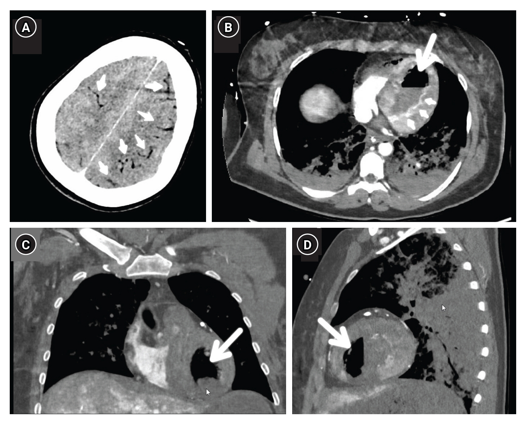
Figure
Reference
-
1. Malik N, Claus PL, Illman JE, Kligerman SJ, Moynagh MR, Levin DL, et al. Air embolism: diagnosis and management. Future Cardiol. 2017; 13:365–78.
Article2. Kandori K, Ishii W, Iiduka R. Massive systemic arterial air embolism caused by an air shunt after blunt chest trauma: a case report. Int J Surg Case Rep. 2018; 51:368–71.
Article3. Ryu SM, Park SM. Unexpected complication during extracorporeal membrane oxygenation support: ventilator associated systemic air embolism. World J Clin Cases. 2018; 6:274–8.
Article4. Shiina G, Shimosegawa Y, Kameyama M, Onuma T. Massive cerebral air embolism following cardiopulmonary resuscitation. Report of two cases. Acta Neurochir (Wien). 1993; 125:181–3.
Article
- Full Text Links
- Actions
-
Cited
- CITED
-
- Close
- Share
- Similar articles
-
- Veno-venous Extracorporeal Membrane Oxygenation with a Double Lumen Catheter for Pediatric Pulmonary Support
- Right ventricular assist device with an oxygenator using extracorporeal membrane oxygenation as a bridge to lung transplantation in a patient with severe respiratory failure and right heart decompensation
- Iatrogenic Iliac Vein Injury Following Extracorporeal Membrane Oxygenation Cannulation in a Patient with May-Thurner Syndrome: A Case Report and Literature Review
- Extracorporeal Membrane Oxygenation for Complicated Scrub Typhus
- Percutaneous bicaval dual lumen cannula for extracorporeal life support


