Ann Clin Neurophysiol.
2024 Apr;26(1):36-37. 10.14253/acn.23016.
Abnormal somatosensory evoked potential findings in syringomyelia
- Affiliations
-
- 1Department of Neurology, Pusan National University Yangsan Hospital, Yangsan, Korea
- 2Department of Neurology, Pusan National University School of Medicine, Yangsan, Korea
- KMID: 2554854
- DOI: http://doi.org/10.14253/acn.23016
Keyword
Figure
-
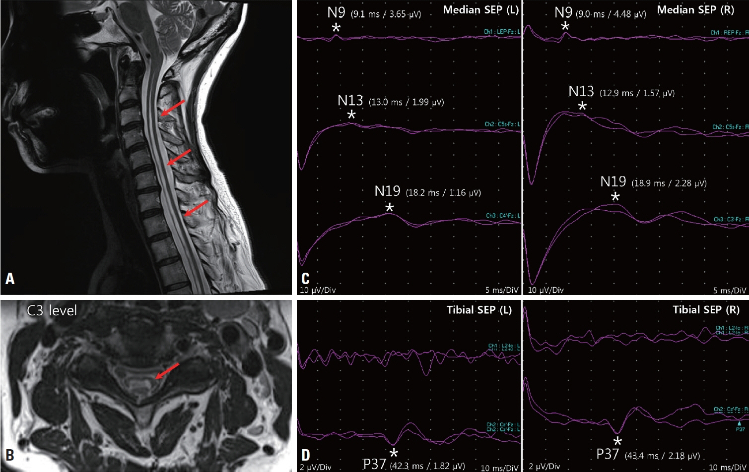
Fig. 1. Sagittal (A) and axial (B, C3 level) images from cervical spine T2-weighted magnetic resonance imaging. An extensive syrinx had developed within the spinal cord (from C2 to T10) (arrows). Median somatosensory evoked potentials (SEPs) (C) were normal for N9 but small for N13 and N19. The tibial SEP (D) was normal for P37 bilaterally in the lower limbs. *Represent the locations of each potential.
Reference
-
1. Giner J, Pérez López C, Hernández B, Gómez de la Riva Á, Isla A, Roda JM. Update on the pathophysiology and management of syringomyelia unrelated to Chiari malformation. Neurologia (Engl Ed). 2019; 34:318–325.2. Morioka T, Kurita-Tashima S, Fujii K, Nakagaki H, Kato M, Fukui M. Somatosensory and spinal evoked potentials in patients with cervical syringomyelia. Neurosurgery. 1992; 30:218–222.3. Anderson NE, Frith RW, Synek VM. Somatosensory evoked potentials in syringomyelia. J Neurol Neurosurg Psychiatry. 1986; 49:1407–1410.
- Full Text Links
-
- Actions
-
Cited
- CITED
-
- Close
- Share
-
- Similar articles
-
- Somatosensory Evoked Potential Study in Patients with Polyneuropathy with Chronic Renal Failure
- The Value of Bulbocavernosus Reflex Latency and Dorsal Nerve Somatosensory Evoked Potential in Diagnosis of Neurogenic Impotence
- Considerations in measuring somatosensory evoked potential
- Evoked Potentials in Wilson Disease
- Clinical Usefulness of Somatosensory Evoked Potentials in Patients with Stroke
