Color discrepancy of single-shade composites at different distances from the interface measured using cell phone images
- Affiliations
-
- 1Graduate Program in Dentistry, Federal University of Sergipe, Aracaju, SE, Brazil
- 2Department of Dentistry, Federal University of Sergipe, Aracaju, SE, Brazil
- KMID: 2554135
- DOI: http://doi.org/10.5395/rde.2024.49.e7
Abstract
Objectives
This study aimed to evaluate the impact of substrate color and interface distance on the color adjustment of 2 single-shade composites, Vittra APS Unique and Charisma Diamond One.
Materials and Methods
Dual disc-shaped specimens were created using Vittra APS Unique or Charisma Diamond One as the center composite, surrounded by shaded composites (A1 or A3). Color measurements were taken with a spectrophotometer against a gray background, recording the color coordinates in the CIELAB color space. Illumination with a lightcorrecting device and image acquisition using a polarizing filter-equipped cell phone were performed on specimens over the same background. Image processing software was used to measure the color coordinates in the center and periphery of the inner composite and in the outer composite. The color data were then converted to CIELAB coordinates and adjusted using data from the spectrophotometer. Color differences (ΔE00 ) between the center/ periphery of single-shade and outer composites were calculated, along with color changes in single-shade composites caused by different outer composites. Color differences for the inner composites surrounded by A1 and A3 were also calculated. Data were analyzed using repeated-measures analysis of variance (α = 0.05).
Results
The results showed that color discrepancies were lowest near the interface and when the outer composite was whiter (A1). Additionally, Charisma Diamond One exhibited better color adjustment ability than Vittra APS Unique.
Conclusions
Color discrepancies between the investigated single-shade composites diminished towards the interface with the surrounding composite, particularly when the latter exhibited a lighter shade.
Keyword
Figure
-
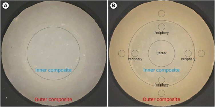
Figure 1 Illustrative specimens’ images, highlighting the delimited areas used for color measurements. (A) An 8-mm diameter area in the center of the specimen was utilized to adjust the values obtained from image measurements with those obtained from the spectrophotometer. (B) Four 1-mm diameter areas were delimited in the outer composite, along with 4 similar areas in the periphery of the inner single-shade composite. Additionally, a centered 4-mm diameter area was also delimited for measurement purposes.
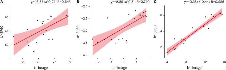
Figure 2 Scatter plots illustrating the color coordinates data of the specimens’ center, which were measured using both the spectrophotometer and the images. Each plot represents a specific color coordinate: (A) L* coordinate, (B) a* coordinate, and (C) b* coordinate. The linear regression equations for each plot are also displayed, depicting the relationships between the measured values obtained from both methods.
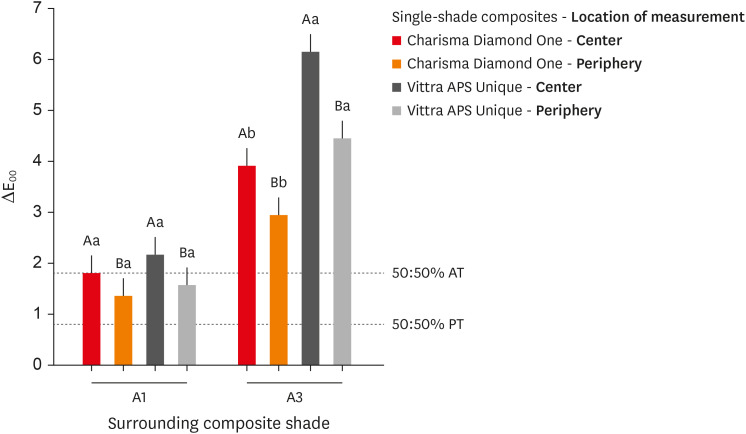
Figure 3 Color differences measured between the inner single-shade composites and the surrounding shaded composite. Letters indicating the statistical differences in Tukey’s test should be analyzed separately for each surrounding composite shade. Uppercase letters compare the locations of evaluation within the same composite, while lowercase letters compare the single-shade composites within the same location. Statistical differences are denoted by distinct letters (p < 0.05).AT, acceptability threshold; PT, perceptibility threshold.
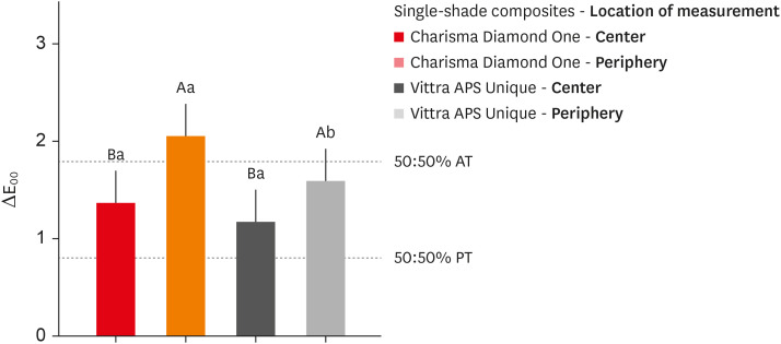
Figure 4 Color differences measured between specimens with outer composite A1 and those with A3 within the same single-shade composite. Uppercase letters compare the locations of evaluation within the same composite, while lowercase letters compare the single-shade composites within the same location. Statistical differences are denoted by distinct letters (p < 0.05).AT, acceptability threshold; PT, perceptibility threshold.
Reference
-
1. Trifkovic B, Powers JM, Paravina RD. Color adjustment potential of resin composites. Clin Oral Investig. 2018; 22:1601–1607.2. Barros MS, Silva PF, Santana ML, Bragança RM, Faria-E-Silva AL. Effect of surrounded shade and specimen’s thickness on color adjustment potential of a single-shade composite. Braz Dent J. 2022; 33:126–132. PMID: 36287494.3. Barros MS, Silva PF, Santana ML, Bragança RM, Faria-E-Silva AL. Effects of surrounding and underlying shades on the color adjustment potential of a single-shade composite used in a thin layer. Restor Dent Endod. 2023; 48:e7. PMID: 36875813.4. de Abreu JL, Sampaio CS, Benalcázar Jalkh EB, Hirata R. Analysis of the color matching of universal resin composites in anterior restorations. J Esthet Restor Dent. 2021; 33:269–276. PMID: 32989879.5. Korkut B, Ünal T, Can E. Two-year retrospective evaluation of monoshade universal composites in direct veneer and diastema closure restorations. J Esthet Restor Dent. 2023; 35:525–537. PMID: 36478098.6. Forabosco E, Consolo U, Mazzitelli C, Kaleci S, Generali L, Checchi V. Effect of bleaching on the color match of single-shade resin composites. J Oral Sci. 2023; 65:232–236. PMID: 37532526.7. Durand LB, Ruiz-López J, Perez BG, Ionescu AM, Carrillo-Pérez F, Ghinea R, et al. Color, lightness, chroma, hue, and translucency adjustment potential of resin composites using CIEDE2000 color difference formula. J Esthet Restor Dent. 2021; 33:836–843. PMID: 33283966.8. Barros MS, Silva PF, Santana ML, Bragança RM, Faria-E-Silva AL. Background and surrounding colors affect the color blending of a single-shade composite. Braz Oral Res. 2023; 37:e035. PMID: 37132724.9. de Melo Oliveira I, Santana TR, Correia AC, Fontes LS, Griza S, Faria-E-Silva AL. Color heterogeneity and individual color changes in dentin and enamel bleached in the presence of a metallic orthodontic bracket. J Esthet Restor Dent. 2021; 33:262–268. PMID: 32955789.10. de Livi GJ, Santana TR, Bragança RM, de Bragança Garcez RM, Faria-E-Silva AL. The role of interface distance and underlying substrate on the color adjustment potential of single-shade composites. J Esthet Restor Dent. 2023; 35:1279–1285. PMID: 37435810.11. Tam WK, Lee HJ. Dental shade matching using a digital camera. J Dent. 2012; 40(Supplement 2):e3–ee10.12. Lazar R, Culic B, Gasparik C, Lazar C, Dudea D. The accuracy of dental shade matching using cross-polarization photography. Int J Comput Dent. 2019; 22:343–351. PMID: 31840142.13. de Bragança RM, Moraes RR, Faria-E-Silva AL. Color assessment of resin composite by using cellphone images compared with a spectrophotometer. Restor Dent Endod. 2021; 46:e23. PMID: 34123759.14. Rondón LF, Ramírez R, Pecho OE. Comparison of visual shade matching and photographic shade analysis. J Esthet Restor Dent. 2022; 34:374–382. PMID: 35128799.15. Brokos I, Polychronakis N, Polyzois G, Lagouvardos P, Krejci I. Illuminant metameric effects on interbrand and intrabrand color differences of direct composite resins. J Prosthet Dent. 2022; 128:1342–1349. PMID: 34045050.16. Yilmaz B, Dede DÖ, Diker E, Fonseca M, Johnston WM, Küçükekenci AS. Effect of cross-polarization filters on the trueness of colors obtained with a single-lens reflex camera, macro lens, and a ring flash. J Esthet Restor Dent. 2023; 35:878–885. PMID: 37073977.17. Soares KD, Bragança RM, Leal PC, Schneider LF, Faria-e-Silva AL. Is it possible to determine the optical properties of resin composites with clinical spectrophotometers? Color Res Appl. 2022; 47:706–716.18. ISO 11664-2:2007. Colorimetry — Part 2: CIE standard illuminants. Geneva: International Organization for Standardization;2007.19. ISO/CIE 11664-1:2019. Colorimetry — Part 1: CIE standard colorimetric observers. Geneva: International Organization for Standardization;2019.20. Araujo FS, Barros MC, Santana ML, de Jesus Oliveira LS, Silva PF, Lima GD, et al. Effects of adhesive used as modeling liquid on the stability of the color and opacity of composites. J Esthet Restor Dent. 2018; 30:427–433. PMID: 29607618.21. Carney MN, Johnston WM. A novel regression model from RGB image data to spectroradiometric correlates optimized for tooth colored shades. J Dent. 2016; 51:45–48. PMID: 27260343.22. Luo MR, Cui BR, Rigg B. The development of the CIE 2000 colour-difference formula: CIEDE2000. Color Res Appl. 2001; 26:340–350.23. Sharma G, Wu W, Dalal EM. The CIEDE2000 color-difference formula: Implementation notes, supplementary test data, and mathematical observations. Color Res Appl. 2005; 30:21–30.24. Paravina RD, Ghinea R, Herrera LJ, Bona AD, Igiel C, Linninger M, et al. Color difference thresholds in dentistry. J Esthet Restor Dent. 2015; 27(Supplement 1):S1–S9. PMID: 25886208.25. Nixon M, Outlaw F, Leung TS. Accurate device-independent colorimetric measurements using smartphones. PLoS One. 2020; 15:e0230561. PMID: 32214340.
- Full Text Links
-
- Actions
-
Cited
- CITED
-
- Close
- Share
-
- Similar articles
-
- Depth-Dependent Performance of Single-Shade Composite Resin: Assessing Color Adjustment Potential and Translucency
- The evaluation of color and color difference according to the layering placement of Incisal shade composites on the body composites of the indirect resin restoration
- Color Matching of Single-Shade Composite Resin by Various Pulp Capping Materials in Anterior Teeth
- Evaluation of the Color Adjustment Potential of Single-Shade Composite Resin in Primary Teeth
- Optical characteristics of resin composite before and after polymerization
