Clin Endosc.
2024 Mar;57(2):268-269. 10.5946/ce.2023.275.
A remnant cystic duct presenting as a duodenal subepithelial tumor
- Affiliations
-
- 1Department of Internal Medicine, Pusan National University School of Medicine and Biomedical Research Institute, Pusan National University Hospital, Busan, Korea
- KMID: 2553765
- DOI: http://doi.org/10.5946/ce.2023.275
Figure
-
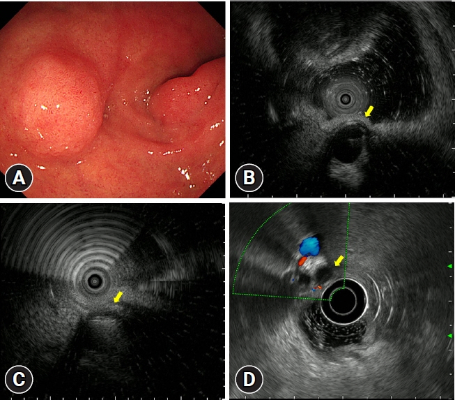
Fig. 1. Endoscopic ultrasonography (EUS) findings. (A) A 1-cm-sized subepithelial lesion covered by normal duodenal mucosa is observed in the anterior wall of the duodenal bulb. (B, C) EUS using a 20-MHz catheter probe reveals a 0.8×0.6 cm-sized anechoic lesion with hyperechoic suture material (arrow) outside the duodenal wall. (D) Conventional EUS (7.5 MHz) reveals that the anechoic lesion was connected to the common bile duct and had no Doppler flow (arrow).
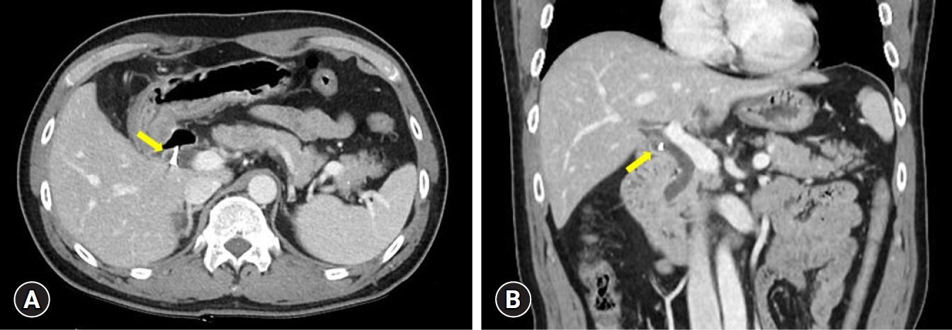
Fig. 2. Abdominal computed tomography reveals a remnant cystic duct with suture material (arrows). (A) Transverse section view. (B) Coronal section view.
Reference
-
1. Eum JS, Kim GH, Park CH, et al. A remnant cystic duct cancer presenting as a duodenal submucosal tumor. Gastrointest Endosc. 2008; 67:975–976.2. Han SY, Chon HK, Kim SH, et al. Quality indicators of endoscopic ultrasound in the pancreatobiliary system: a brief review of current guidelines. Clin Endosc. 2024; 57:158–163.3. Ryou SH, Kim HJ. Successful removal of remnant cystic duct stump stone using single-operator cholangioscopy-guided electrohydraulic lithotripsy: two case reports. Clin Endosc. 2023; 56:375–380.
- Full Text Links
-
- Actions
-
Cited
- CITED
-
- Close
- Share
-
- Similar articles
-
- An Incidentally Detected Remnant Cystic Duct Carcinoma duringthe Evaluation of a Duodenal Submucosal Tumor (SMT) Lesion
- Clinical Significance of Extraluminal Compressions according to the Site of the Duodenum
- Laparoscopic Resection for the Treatment of Symptomatic Remnant Huge Cystic Duct with Stone after Laparoscopic Cholecystectomy
- Intraductal Papillary Mucinous Neoplasm with Associated Invasive Carcinoma Arising from Remnant Cystic Duct: A Case Report
- Laparoscopic Removal of a Retained Gallbladder with Remnant Cystic Duct Calculi
