A Case of Primary Hepatic Leiomyoma Incidentally Found in a Middle-aged Woman without Underlying Diseases
- Affiliations
-
- 1Department of Surgery, Inje University Haeundae Paik Hospital, Inje University College of Medicine, Busan, Korea
- KMID: 2552242
- DOI: http://doi.org/10.4166/kjg.2023.143
Figure
-

Fig. 1 Initial dynamic CT scan. (A, B) Abdominal CT scan demonstrating a 6.3 cm progressive enhancing mass in S7 (arrow) of the liver in portal phase (A) and venous phase (B).

Fig. 2 Dynamic MRI findings. (A) Hypointense signal on T1-weighted image. (B) Hyperintense signal on T2-weighted image. (C) Heterogeneously enhancement in the arterial phase. (D) Hypointense signal in hepatobiliary delayed phase.
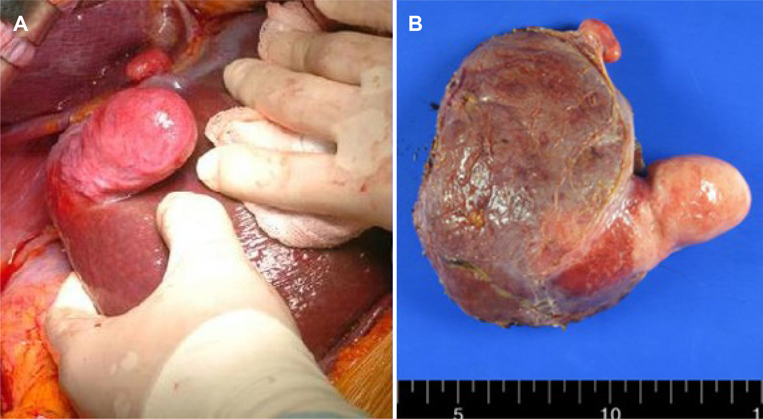
Fig. 3 Gross pathologic findings. (A) Exophytic mass on S7 in operative field. (B) Gross finding of the tumor shows grayish white, solid, and whorled mass weighing 171 g and 7.5×4.2×4.0 cm.

Fig. 4 Hematoxylin and eosin stain, x100. The hepatic tumor was composed of bundles of spindle-shaped cells with nuclei and eosinophilic cytoplasm.

Fig. 5 Immunohistochemical examination. (A–D) Smooth muscle actin, x100 (A), KIT, x100 (B), CD34, x100 (C), and S100, x100 (D) were examined by immunohistochemistry.

Fig. 6 CT scan at postoperative 30th day. There were no abnormal findings except for small amount fluid collection at the resection margin.
Reference
-
1. Reinertson TE, Fortune JB, Peters JC, Pagnotta I, Balint JA. 1992; Primary leiomyoma of the liver. A case report and review of the literature. Dig Dis Sci. 37:622–627. DOI: 10.1007/BF01307591. PMID: 1551357.2. Luo XZ, Ming CS, Chen XP, Gong NQ. 2013; Epstein-Barr virus negative primary hepatic leiomyoma: case report and literature review. World J Gastroenterol. 19:4094–4098. DOI: 10.3748/wjg.v19.i25.4094. PMID: 23840159. PMCID: PMC3703201.3. Demel R. 1926; Ein operierter Fall von Leber-Myom. Virchows Arch Pathol Anat Physiol Klin Med. 261:881–884. DOI: 10.1007/BF01892215.4. Prévot S, Néris J, de Saint Maur PP. 1994; Detection of Epstein Barr virus in an hepatic leiomyomatous neoplasm in an adult human immunodeficiency virus 1-infected patient. Virchows Arch. 425:321–325. DOI: 10.1007/BF00196156. PMID: 7812519.5. Hollands MJ, Jaworski R, Wong KP, Little JM. 1989; A leiomyoma of the liver. HPB Surg. 1:337–343. DOI: 10.1155/1989/45978. PMID: 2487073. PMCID: PMC2423549.6. Belli G, Ciciliano F, Lannelli A, Marano I. 2001; Hepatic resection for primary giant leiomyoma of the liver. HPB (Oxford). 3:11–12. DOI: 10.1080/136518201753173692. PMID: 18333008. PMCID: PMC2020611.7. Santos I, Valls C, Leiva D, Serrano T, Martinez L, Ruiz S. 2011; Primary hepatic leiomyoma: case report. Abdom Imaging. 36:315–317. DOI: 10.1007/s00261-010-9648-y. PMID: 20959979.8. Penn I. 1978; Malignancies associated with immunosuppressive or cytotoxic therapy. Surgery. 83:492–502.9. Lee ES, Locker J, Nalesnik M, et al. 1995; The association of Epstein-Barr virus with smooth-muscle tumors occurring after organ transplantation. N Engl J Med. 332:19–25. DOI: 10.1056/NEJM199501053320104. PMID: 7990861.10. Starzl TE, Nalesnik MA, Porter KA, et al. 1984; Reversibility of lymphomas and lymphoproliferative lesions developing under cyclosporin-steroid therapy. Lancet. 1:583–587. DOI: 10.1016/S0140-6736(84)90994-2. PMID: 6142304.11. Jang SH, Kim SM, Sohn JS, Ryu KH, Yuk HB. 2016; A firm hepatic mass cannot be penetrated by US-guided needle biopsy. Clin Ultrasound. 1:126–129. DOI: 10.18525/cu.2016.1.2.126.
