J Yeungnam Med Sci.
2024 Jan;41(1):45-47. 10.12701/jyms.2023.01018.
Sciatic neurotmesis and periostitis ossificans progressiva due to a traumatic/unexpected glass injury: a case report
- Affiliations
-
- 1Department of Physical Medicine and Rehabilitation, Hacettepe University Medical School, Ankara, Turkiye
- KMID: 2551333
- DOI: http://doi.org/10.12701/jyms.2023.01018
Abstract
- Peripheral nerves may be affected or injured for several reasons. Peripheral nerve damage can result from trauma, surgery, anatomical abnormalities, entrapment, systemic diseases, or iatrogenic injuries. Trauma and iatrogenic injuries are the most common causes. The ulnar, median, and radial nerves are the most injured nerves in the upper extremities, while the sciatic and peroneal nerves are the most injured nerves in the lower extremities. The clinical symptoms of peripheral nerve damage include pain, weakness, numbness/tingling, and paresthesia. Therefore, early diagnosis and appropriate treatment of peripheral nerve injuries are crucial. If a peripheral nerve injury is left untreated, it can lead to severe complications and significant morbidity. The sciatic nerve is one of the most affected nerves. This nerve is generally injured by trauma and iatrogenic causes. Children are more susceptible to trauma than adults. Therefore, sciatic nerve injuries are observed in pediatric patients. When the sciatic nerve is damaged, pain, weakness, sensory loss, and gait disturbances can occur. Therefore, the diagnosis and treatment of sciatic nerve injuries are important to avoid unexpected consequences. Ultrasound can play an important role in the diagnosis of peripheral nerve injury and the follow-up of patients. The aim of this case report is twofold. First, we aimed to emphasize the critical role of ultrasonographic evaluation in the diagnosis of peripheral nerve injuries and pathologies. Second, we aimed to present this case, which has distinguishing features, such as the existence of periostitis ossificans progressiva with sciatic neurotmesis due to a traumatic glass injury.
Keyword
Figure
-
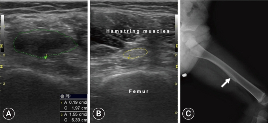
Fig. 1. (A) Axial imaging for cross-sectional area measurement (1.55 cm2) shows the edematous, irregular, and anechoic sciatic nerve (green tracing) on the left side. (B) Axial imaging for cross-sectional area measurement (0.19 cm2) shows the normal sciatic nerve (yellow tracing) on the right side. (C) Anteroposterior radiograph of the left femur (frog leg position) shows the periosteal lesion (arrow).
Reference
-
References
1. Costales JR, Socolovsky M, Sánchez Lázaro JA, Álvarez García R. Peripheral nerve injuries in the pediatric population: a review of the literature. Part I: traumatic nerve injuries. Childs Nerv Syst. 2019; 35:29–35.2. Mc Donald CM. Electrodiagnosis in pediatrics. In : Alexander MA, Matthews DJ, editors. Pediatric rehabilitation: principles and practice. 5th ed. New York: Demos Medical Publishing;2015. p. 113–52.3. Jaque-Almendras C, Escobar RG, Caicedo-Feijoo A, Beytía-Reyes ML, Correa-Pérez S, Gejman-Enríquez R, et al. Pediatric sciatic neuropathy: clinical presentation and long term follow up. Rev Chil Pediatr. 2020; 91:85–93.4. Chang KV, Şahin Onat Ş, Lee CW, Kara M, Hung CY, Özçakar L. EURO-MUSCULUS/USPRM basic scanning protocols revisited in children. Eur J Phys Rehabil Med. 2016; 52:887–901.5. Hung CY, Hsiao MY, Özçakar L, Chang KV, Wu CH, Wang TG, et al. Sonographic tracking of the lower limb peripheral nerves: a pictorial essay and video demonstration. Am J Phys Med Rehabil. 2016; 95:698–708.6. Ozçakar L, Carli AB, Tok F, Tekin L, Akkaya N, Kara M. The utility of musculoskeletal ultrasound in rehabilitation settings. Am J Phys Med Rehabil. 2013; 92:805–17.
