Clin Endosc.
2024 Jan;57(1):137-139. 10.5946/ce.2023.170.
Nasopharyngeal examination during transoral upper gastrointestinal endoscopy
- Chong VH
 1,2
1,2
- Affiliations
-
- 1Department of Internal Medicine, Raja Isteri Pengiran Anak Saleha Hospital, Bandar Seri Begawan, Brunei Darussalam
- 2Department of Medicine, PMMPHMAB Hospital, Tutong, Brunei Darussalam
- KMID: 2551205
- DOI: http://doi.org/10.5946/ce.2023.170
Figure
-
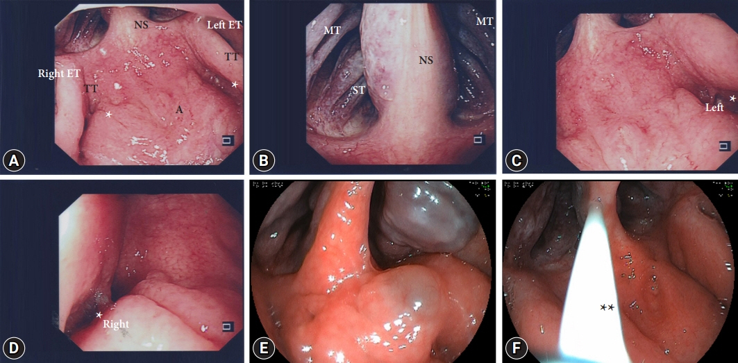
Fig. 1. Retroflexed view of the nasopharynx. (A–D) Normal. (E) A patient with adenoid cyst and congested left superior turbinate. (F) A patient with mucus discharge (**). Images are inverted; top and bottom, and left and right. A adenoid pad or roof of nasopharynx; NS, nasal septum; TT, torus tubaris or torus of the auditory tube; ET, eustachian tube; *, fossa of Rosenmuller; MT, middle turbinate; ST, superior turbinate.
Reference
-
1. Ono Y, Yao K, Takaki Y, et al. Efficacy of endoscopy under general anesthesia for the detection of synchronous lesions in oro-hypopharyngeal cancer. Clin Endosc. 2023; 56:315–324.2. Noh JH, Kim DH. Endoscopy under general anesthesia for detecting synchronous lesions of head and neck squamous cell carcinoma. Clin Endosc. 2023; 56:308–309.3. World Cancer Research Fund International. Nasopharyngeal cancer statistics [Internet]. London: World Cancer Research Fund International;2020 [cited 2023 Jun 29]. Available from: https://www.wcrf.org/cancer-trends/nasopharyngeal-cancer-statistics/.4. World Cancer Research Fund International. Mouth and oral cancer statistics [Internet]. London: World Cancer Research Fund International;2020 [cited 2023 Jun 29]. Available from https://www.wcrf.org/cancer-trends/mouth-and-oral-cancer-statistics/.
- Full Text Links
-
- Actions
-
Cited
- CITED
-
- Close
- Share
-
- Similar articles
-
- Transoral Adenoidectomy with the Microdebrider under Transnasal Endoscopy
- Introduction to Starting Upper Gastrointestinal Endoscopy: Proper Insertion, Complete Observation, and Appropriate Photographing
- Upper gastrointestinal diseases diagnosed by upper gastrointestinal fiberoptic endoscopy in children
- Observable Laryngopharyngeal Lesions during the Upper Gastrointestinal Endoscopy
- A Study of the Upper Gastrointestinal Polyp
