Acute Crit Care.
2023 Nov;38(4):513-514. 10.4266/acc.2023.01228.
Right-sided infective endocarditis of a native valve with multiple embolus lesions
- Affiliations
-
- 1Department of Internal Medicine, Jeju National University College of Medicine, Jeju, Korea
- KMID: 2550909
- DOI: http://doi.org/10.4266/acc.2023.01228
Figure
-
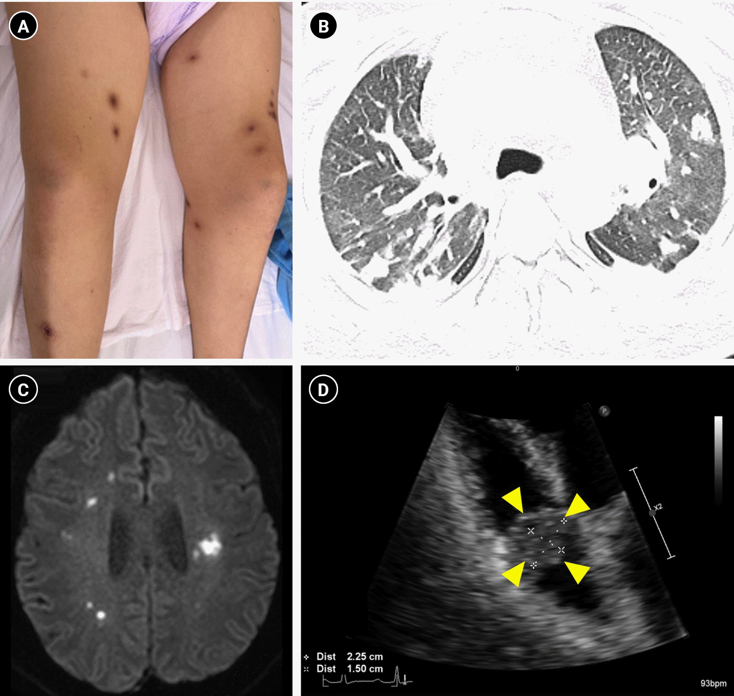
Figure 1. (A) Multiple dot-like black erythematous lesions measuring 1–3 cm throughout the body. (B) Chest computed tomography revealing multiple nodules in both lungs. (C) Diffusion-weighted brain magnetic resonance imaging of lesions with high signal intensity, appearing as multiple scattered dots, in both fronto-parieto-temporal lobes. (D) Apical view of the four chambers on transthoracic echocardiography showing vegetation in the tricuspid valve indicated by yellow arrowheads.
Reference
-
1. Shmueli H, Thomas F, Flint N, Setia G, Janjic A, Siegel RJ. Right-sided infective endocarditis 2020: challenges and updates in diagnosis and treatment. J Am Heart Assoc. 2020; 9:e017293.
Article2. Habib G, Lancellotti P, Antunes MJ, Bongiorni MG, Casalta JP, Del Zotti F, ESC Scientific Document Group, et al. 2015 ESC Guidelines for the management of infective endocarditis: The Task Force for the Management of Infective Endocarditis of the European Society of Cardiology (ESC). Endorsed by: European Association for Cardio-Thoracic Surgery (EACTS), the European Association of Nuclear Medicine (EANM). Eur Heart J. 2015; 36:3075–128.
Article
- Full Text Links
-
- Actions
-
Cited
- CITED
-
- Close
- Share
-
- Similar articles
-
- A case of Haemophilus aphrophilus native valve endocarditis
- Single and Multiple Valve Surgery in Native Valve Infective Endocarditis
- A case of native valve infective endocarditis caused by Microbacterium species
- Active Infective Endocarditis with Vegetation of Right Atrium in Patient with End-stage Renal Disease
- A Case of Infective Endocarditis Complicated with Multiple Myocotic Aneurysm and Mitral Valve Perforation
