Extensive Papillary Muscle Calcification in Adult Anomalous Left Coronary Artery from Pulmonary Artery
- Affiliations
-
- 1Department of Cardiology, Sree Chitra Tirunal Institute for Medical Science and Technology (SCTIMST), Trivandrum, Kerala, India
- 2Department of Imaging Sciences & Intervention Radiology, Sree Chitra Tirunal Institute for Medical Sciences and Technology (SCTIMST), Trivandrum, Kerala, India
- KMID: 2550610
- DOI: http://doi.org/10.4070/kcj.2023.0243
Figure
-
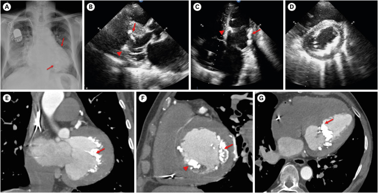
Figure 1 (A) Chest X-ray showing opacity in cardiac silhouette raising suspicion of calcification of mediastinal structure; 2-dimensional-echocardiogram in parasternal long axis (B), apical 4-chamber (C) and parasternal short axis (D) views showing extensive anterolateral (arrow) and posteromedial (arrowhead) papillary muscle with severe endocardial (D) calcification having acoustic shadow; Cardiac computed tomogram in coronal (E), sagittal (F) and axial (G) view showing extensive anterolateral (arrow) and posteromedial (arrowhead) papillary muscle with severe endocardial (D) calcification.

Figure 2 Volume rendering image showing extensive myocardial calcification (note that calcification is restricted to left coronary territory without affecting right coronary territory). Note the stump of left main coronary artery which was ligated during surgery (arrowhead). Asterix: myocardial calcification with likely both papillary muscles calcification.LAD = left anterior descending artery; LIMA = left internal mammary artery; RCA = right coronary artery.
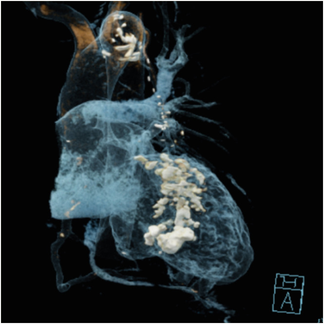
Figure 3 Cinematic rendering of the heart with window level adjusted for high attenuation structures showing dense calcifications in the left ventricle. White colour: calcification.
Reference
-
1. Mondal S, Raj RR, Harikrishnan S. Calcium jacket of left ventricle: an aristocracy of the monster. Indian J Clin Cardiol. 2023; 4:204–207.2. Nance JW Jr, Crane GM, Halushka MK, Fishman EK, Zimmerman SL. Myocardial calcifications: pathophysiology, etiologies, differential diagnoses, and imaging findings. J Cardiovasc Comput Tomogr. 2015; 9:58–67. PMID: 25456525.3. Math RS, Parakh N, Sarin SS, Tyagi S. Anomalous origin of the left coronary artery from the pulmonary artery (ALCAPA) presenting as a complete heart block. Pediatr Cardiol. 2010; 31:526–529. PMID: 20165845.4. Sivasubramonian S, Bohora S. Serpigenous calcification in the heart. Pediatr Cardiol. 2009; 30:557–559. PMID: 19294458.
- Full Text Links
-
- Actions
-
Cited
- CITED
-
- Close
- Share
-
- Similar articles
-
- Anomalous origin of the left coronary artery from the pulmonary artery
- Congenital Absence of Left Circumflex Coronary Artery: Circumflex Artery Extended from Right Coronary Artery
- Sudden Death Associated with Anomalous Left Coronary Artery Origin from Right Sinus of Valsalva with Posterior Course
- Anomalous Origin of Left Coronary Artery from Pulmonary Artery:Report of an Adult Case
- Anomalous Origin of the Left Coronary Artery from the Pulmonary Artery in an Adult: A case report
