Bone remodeling of the fibula segment as a form of neocondyle after free vascularized bone transfer: a report of two cases
- Affiliations
-
- 1Department of Oral and Maxillofacial Surgery, Asan Medical Center, College of Medicine, University of Ulsan, Seoul, Korea
- KMID: 2550299
- DOI: http://doi.org/10.5125/jkaoms.2023.49.6.354
Abstract
- The temporomandibular joint is a unique structure composed of the joint capsule, articular disc, mandibular condyles, glenoid fossa of the temporal bone, surrounding ligaments, and associated muscles. The condyle is one of the major components of a functional temporomandibular joint. Reconstruction of large mandibular defects involving the condyle is a surgical challenge for oral and maxillofacial surgeons. To restore large mandibular defects, there are different options for free flap method such as fibula, scapula, and iliac crest. Currently, the vascularized fibula free flap is the gold standard for reconstruction of complex mandibular defects involving the condyle. In the present report, neocondyle regeneration after mandible reconstruction including the condyle head with fibula free flap was evaluated. In this report, two patients were evaluated periodically, and remodeling of the distal end of the free fibula was observed in both cases after condylectomy or mandibulectomy. With preservation of the articular disc, trapezoidal shaping of the neocondyle, and elastic guidance of occlusion, neocondyle bone regeneration occured without ankylosis. Preservation of the articular disc and maintenance of proper occlusion are critical factors in regeneration of the neocondyle after mandible reconstruction.
Figure
-

Fig. 1 On the day 6 postoperative panoramic radiograph, the vascularized fibula free flap was fixated with two L-shaped semi-rigid mini plates and three four-hole mini plates.

Fig. 2 The fibula bone was prepared in an L-shape according to the three-dimensional simulation surgical template.
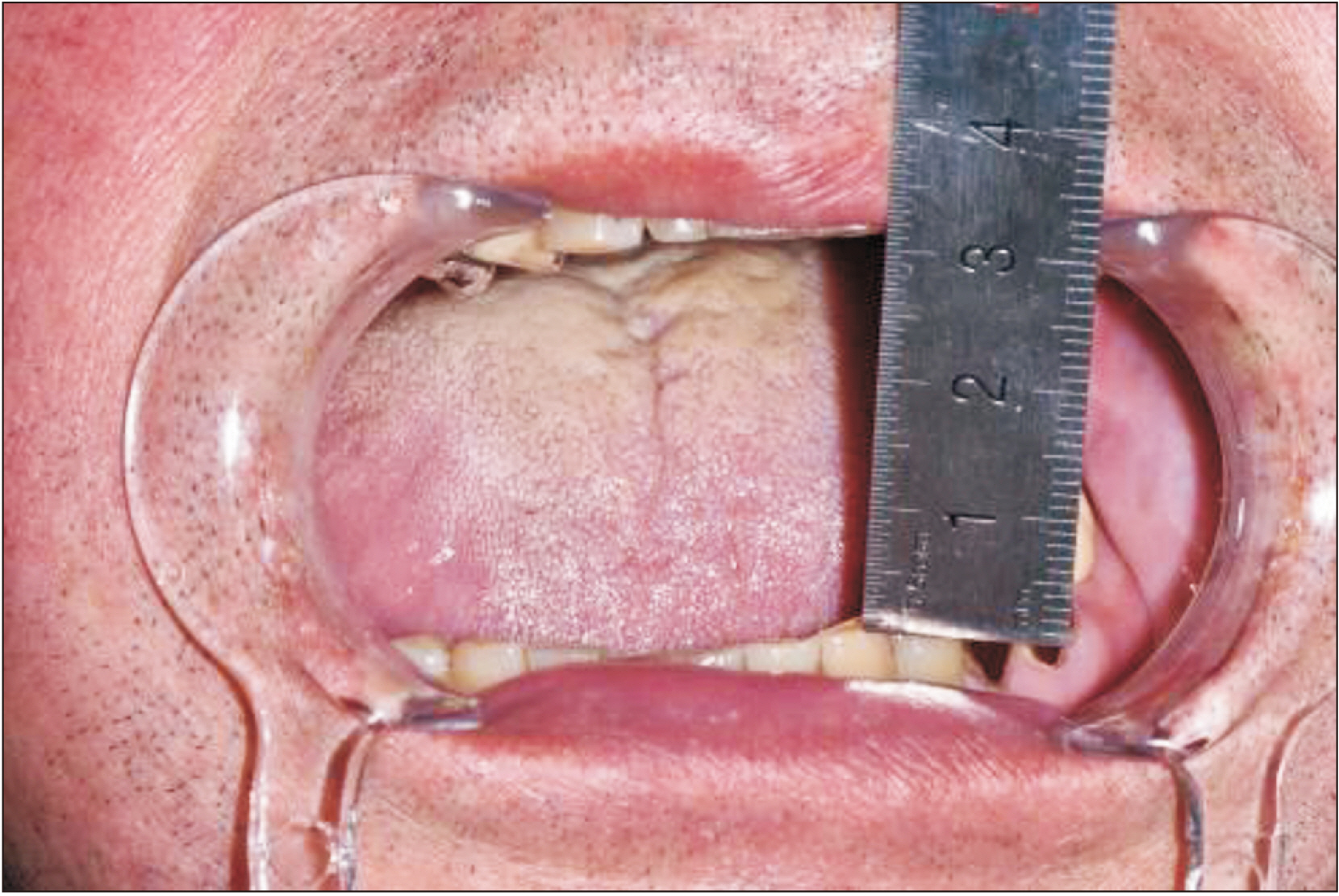
Fig. 3 At the 40-month follow-up, the patient showed 35-mm maximum mouth opening.
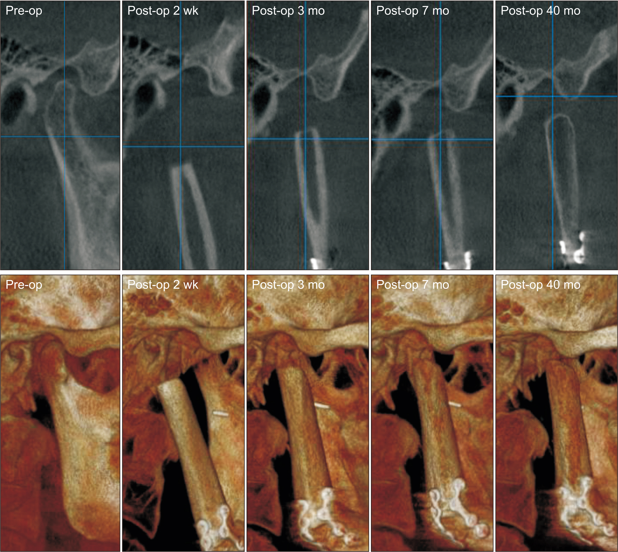
Fig. 4 Rounding off at the distal end of the fibula was observed at the three-month postoperative follow-up.

Fig. 5 The distal end of the fibula was prepared in a right trapezoidal shape using a surgical template.
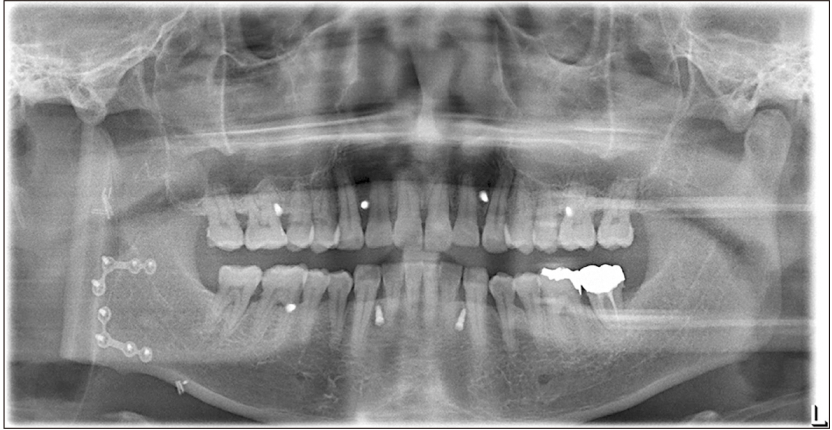
Fig. 6 On the day 4 postoperative panoramic radiograph, the vascularized fibula free flap was fixed with two L-shaped semi-rigid mini plates on the native mandible.

Fig. 7 Neocondyle bone growth in the direction of the lateral pterygoid traction started two months after surgery.
Reference
-
References
1. Schmelzeisen R, Voss PJ, Neukam FW. Greenberg AM, Schmelzeisen R, editors. 2019. Microvascular reconstruction of the condyle and the ascending ramus. Craniomaxillofacial reconstructive and corrective bone surgery. 2nd ed. Springer;p. 427–47. DOI: 10.1007/978-1-4939-1529-3_33.2. Lee ZH, Avraham T, Monaco C, Patel AA, Hirsch DL, Levine JP. 2018; Optimizing functional outcomes in mandibular condyle reconstruction with the free fibula flap using computer-aided design and manufacturing technology. J Oral Maxillofac Surg. 76:1098–106. https://doi.org/10.1016/j.joms.2017.11.008. DOI: 10.1016/j.joms.2017.11.008. PMID: 29222966.3. Yu Y, Zhang WB, Liu XJ, Guo CB, Yu GY, Peng X. 2020; Regeneration of the neocondyle after free fibular flap reconstruction of the mandibular condyle. J Oral Maxillofac Surg. 78:479–87. https://doi.org/10.1016/j.joms.2019.11.009. DOI: 10.1016/j.joms.2019.11.009. PMID: 31838093.4. Yu Y, Soh HY, Bai S, Zhang WB, Wang Y, Peng X. 2021; Three-dimensional morphological analysis of neocondyle bone growth after fibula free flap reconstruction. Int J Oral Maxillofac Surg. 50:1429–34. https://doi.org/10.1016/j.ijom.2021.03.005. DOI: 10.1016/j.ijom.2021.03.005. PMID: 33752937.5. Peled M, El-Naaj IA, Lipin Y, Ardekian L. 2005; The use of free fibular flap for functional mandibular reconstruction. J Oral Maxillofac Surg. 63:220–4. https://doi.org/10.1016/j.joms.2004.06.052. DOI: 10.1016/j.joms.2004.06.052. PMID: 15690291.6. Chaine A, Pitak-Arnnop P, Hivelin M, Dhanuthai K, Bertrand JC, Bertolus C. 2009; Postoperative complications of fibular free flaps in mandibular reconstruction: an analysis of 25 consecutive cases. Oral Surg Oral Med Oral Pathol Oral Radiol Endod. 108:488–95. https://doi.org/10.1016/j.tripleo.2009.05.043. DOI: 10.1016/j.tripleo.2009.05.043. PMID: 19699114.7. Wax MK, Winslow CP, Hansen J, MacKenzie D, Cohen J, Andersen P, et al. 2000; A retrospective analysis of temporomandibular joint reconstruction with free fibula microvascular flap. Laryngoscope. 110:977–81. https://doi.org/10.1097/00005537-200006000-00018. DOI: 10.1097/00005537-200006000-00018. PMID: 10852517.8. Wei H, Pinting L, Enyi T, Zhiyong W. 2012; Initial finding of mandible mass in multiple myeloma. J Craniofac Surg. 23:e599–600. https://doi.org/10.1097/scs.0b013e31826bf44c. DOI: 10.1097/SCS.0b013e31826bf44c.9. Neville BW, Damm DD, Allen CM, Chi AC. Neville BW, Damm DD, Allen CM, Chi AC, editors. 2016. Hematologic disorders. Oral and maxillofacial pathology. 4th ed. Elsevier;p. 563–5.10. Boffano P, Viterbo S, Barreca A, Berrone S. 2011; Pathologic mandibular fracture as the presenting manifestation of multiple myeloma. J Craniofac Surg. 22:1312–5. https://doi.org/10.1097/scs.0b013e31821c6cbe. DOI: 10.1097/SCS.0b013e31821c6cbe. PMID: 21772196.11. Gerbino G, Zavattero E, Bosco G, Berrone S, Ramieri G. 2017; Temporomandibular joint reconstruction with stock and custom-made devices: indications and results of a 14-year experience. J Craniomaxillofac Surg. 45:1710–5. https://doi.org/10.1016/j.jcms.2017.07.011. DOI: 10.1016/j.jcms.2017.07.011. PMID: 28843402.12. Amarista FJ, Jones JP, Brown Z, Rushing DC, Jeske NA, Perez DE. 2022; Outcomes of total joint alloplastic reconstruction in TMJ ankylosis. Oral Surg Oral Med Oral Pathol Oral Radiol. 134:135–42. https://doi.org/10.1016/j.oooo.2021.12.121. DOI: 10.1016/j.oooo.2021.12.121. PMID: 35431176.13. Kang SH, Lee S, Nam W. 2019; Condyle dislocation following mandibular reconstruction using a fibula free flap: complication cases. Maxillofac Plast Reconstr Surg. 41:14. https://doi.org/10.1186/s40902-019-0197-1. DOI: 10.1186/s40902-019-0197-1. PMID: 30997360. PMCID: PMC6441667.14. Meng FW, Zhao JL, Hu KJ, Liu YP. 2009; A new hypothesis of mechanisms of traumatic ankylosis of temporomandibular joint. Med Hypotheses. 73:92–3. https://doi.org/10.1016/j.mehy.2009.01.024. DOI: 10.1016/j.mehy.2009.01.024. PMID: 19261390.15. Liu CK, Liu P, Meng FW, Deng BL, Xue Y, Mao TQ, et al. 2012; The role of the lateral pterygoid muscle in the sagittal fracture of mandibular condyle (SFMC) healing process. Br J Oral Maxillofac Surg. 50:356–60. https://doi.org/10.1016/j.bjoms.2011.05.015. DOI: 10.1016/j.bjoms.2011.05.015. PMID: 21802803.16. Callahan N, Patel M, Dyalram D, Lubek JE. 2022; Is the prevention of condylar sag with maxillomandibular fixation the key to functional reconstruction of a mandibular disarticulation resection? Oral Surg Oral Med Oral Pathol Oral Radiol. 134:317–22. https://doi.org/10.1016/j.oooo.2022.02.012. DOI: 10.1016/j.oooo.2022.02.012. PMID: 35428599.17. Engroff SL. 2005; Fibula flap reconstruction of the condyle in disarticulation resections of the mandible: a case report and review of the technique. Oral Surg Oral Med Oral Pathol Oral Radiol Endod. 100:661–5. https://doi.org/10.1016/j.tripleo.2005.03.016. DOI: 10.1016/j.tripleo.2005.03.016. PMID: 16301145.18. Guyot L, Richard O, Layoun W, Cheynet F, Bellot-Samson V, Chossegros C, et al. 2004; Long-term radiological findings following reconstruction of the condyle with fibular free flaps. J Craniomaxillofac Surg. 32:98–102. https://doi.org/10.1016/j.jcms.2003.11.003. DOI: 10.1016/j.jcms.2003.11.003. PMID: 14980591.19. Wu D, Yang XJ, Cheng P, Deng TG, Jiang X, Liu P, et al. 2015; The lateral pterygoid muscle affects reconstruction of the condyle in the sagittal fracture healing process: a histological study. Int J Oral Maxillofac Surg. 44:1010–5. https://doi.org/10.1016/j.ijom.2015.02.004. DOI: 10.1016/j.ijom.2015.02.004. PMID: 25752241.20. Deng TG, Liu CK, Liu P, Zhang LL, Wu LG, Zhou HZ, et al. 2016; Influence of the lateral pterygoid muscle on traumatic temporomandibular joint bony ankylosis. BMC Oral Health. 16:62. https://doi.org/10.1186/s12903-016-0220-1. DOI: 10.1186/s12903-016-0220-1. PMID: 27234304. PMCID: PMC4884350.
- Full Text Links
-
- Actions
-
Cited
- CITED
-
- Close
- Share
-
- Similar articles
-
- A Clinical Study of Vascularized Osteocutaneous Fibular Transfer
- Treatment of Bone Tumor with Free Vascularized Bone Graft - Comparision with Devascularized Bone Graft -
- Vascularized Fibula Graft for Restoration of the Large Bone Defect
- Vascularized Osteocutaneous Fibular Transfer
- Free Vascularized Osteocutaneous Fibular Graft To The Tibia
