Korean Circ J.
2023 Dec;53(12):858-859. 10.4070/kcj.2023.0240.
A Rare Epicardial Cytokeratin-Positive Interstitial Reticulum Cell Tumor
- Affiliations
-
- 1Department of Radiology, Faculty of Medicine, Kagawa University, Kagawa, Japan
- KMID: 2548794
- DOI: http://doi.org/10.4070/kcj.2023.0240
Figure
-
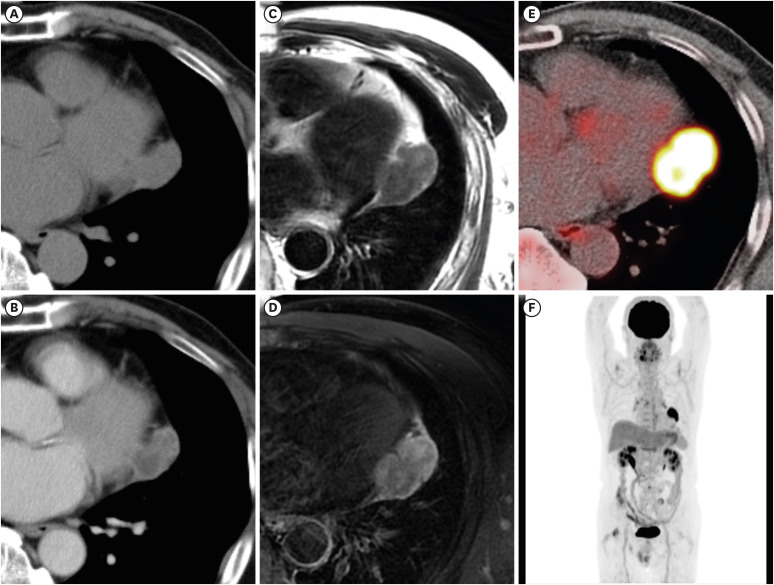
Figure 1 Multimodality imaging in a 71-year-old male. Axial plane nonenhanced (A) and contrast enhanced (B) CT images revealed a poorly enhanced mass around the heart. Magnetic resonance imaging indicated a mass arising from the pericardium of the left ventricle, that appeared mildly hyperintense on T2-weighted images (C) and hyperintense on T2-weighted fat-saturated images (D). FDG positron emission tomography/CT revealed high FDG uptake within the mass (SUVmax: 13.77) (E), while no other abnormal uptakes were observed in the body (F).CT = computed tomography; FDG =18F-fluorodeoxyglucose; SUVmax = maximum standard unit value.
Reference
-
1. Roy S, Das I, Saha K, Roy R, Bhattacharya S. A rare case of cytokeratin-positive interstitial reticulum cell sarcoma and review of the entity. Indian J Surg Oncol. 2015; 6:271–275. PMID: 27217677.2. Kaji S, Hiruta N, Sasai D, Nagashima M, Ohe R, Yamakawa M. Cytokeratin-positive interstitial reticulum cell (CIRC) tumor in the lymph node: a case report of the transformation from the epithelioid cell type to the spindle cell type. Diagn Pathol. 2020; 15:121. PMID: 32979929.3. Dong YC, Wu B, Sheng Z, Wang JD, Zhou HB, Zhou XJ. Cytokeratin-positive interstitial reticulum cell tumors of lymph nodes: a case report and review of literature. Chin Med J (Engl). 2008; 121:658–663. PMID: 18466689.
- Full Text Links
-
- Actions
-
Cited
- CITED
-
- Close
- Share
-
- Similar articles
-
- Renomedullary Interstitial Cell Tumor
- Interdigitating Reticulum Cell Sarcoma of Lymph Node
- A Case of Huge Reticulum Cell Sarcoma of the Brain
- Interdigitating Reticulum Cell Sarcoma of Lymph Node
- Expressions of p53, Ki-67, PCNA and cytokeratin 17, cytokeratin 18 in recurred and non-recurred ameloblastoma
