Primary Cavernous Hemangioma of the Thyroid Gland
- Affiliations
-
- 1Department of Otolaryngology, Kangwon National University Hospital, Chuncheon, Korea
- 2Pathology Center, Seegene Medical Foundation, Seoul, Korea
- 3Department of Otolaryngology, Kangwon National University College of Medicine, Chuncheon, Korea
- KMID: 2548750
- DOI: http://doi.org/10.11106/ijt.2023.16.2.205
Abstract
- Hemangiomas are benign vascular tumors that result from the abnormal proliferation of vascular tissue. Thyroid hemangiomas can develop as a result of procedures such as fine needle aspiration or other secondary trauma. Primary thyroid cavernous hemangioma is an extremely rare condition, with only a few reported cases. In this report, we present the case of an 83-year-old woman who complained of progressively worsening symptoms of right neck obstruction. She was undergoing levothyroxine treatment for hypothyroidism, and there was no specific family history of thyroid issues. The patient presented with a goiter and obstructive symptoms, and denied any history of trauma or invasive procedures. Thyroid sonography revealed a 6.21 cm heterogeneous dominant solid nodule in the right lobe. Additionally, a large mixed cystic and 6 cm solid mass was identified in the right lobe on CT scan. Due to the significant size of the mass and the presence of obstructive symptoms, the patient underwent a right thyroid lobectomy without further evaluations. Histologic examination of the specimen revealed a cavernous thyroid hemangioma. This case report presents our experience in diagnosing cavernous thyroid hemangioma.
Keyword
Figure
-

Fig. 1 Right thyroid ultrasonography (US). (A) Thyroid US (2019) the mass size 4.20×4.56 cm. (B) Thyroid US (2020) the mass size increased at 4.72×5.1 cm. (C) Thyroid US (2022) the mass size increased at 4.98×6.21 cm. All US images consistently depict the mild hypo- and iso-echogenicity, predominantly solid, parallel, and smooth margin; K-TRIADS 3.
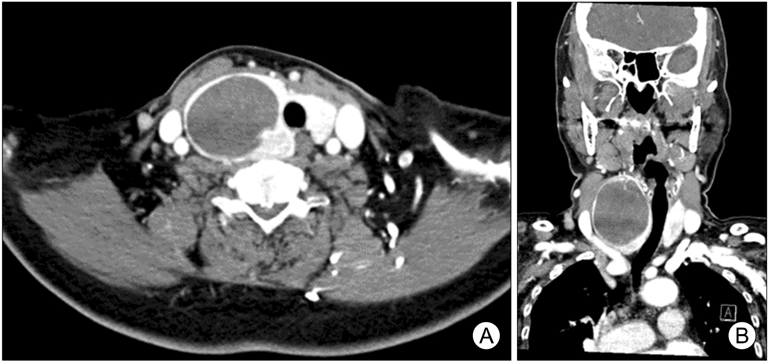
Fig. 2 Enhanced thyroid CT (left: axial, right: coronal). (A) Large mixed cystic and solid mass in right thyroid lobe around 6 cm in axial view. (B) Well defined heterogeneously enhancing large mass and showing tracheal compression and shift to left.
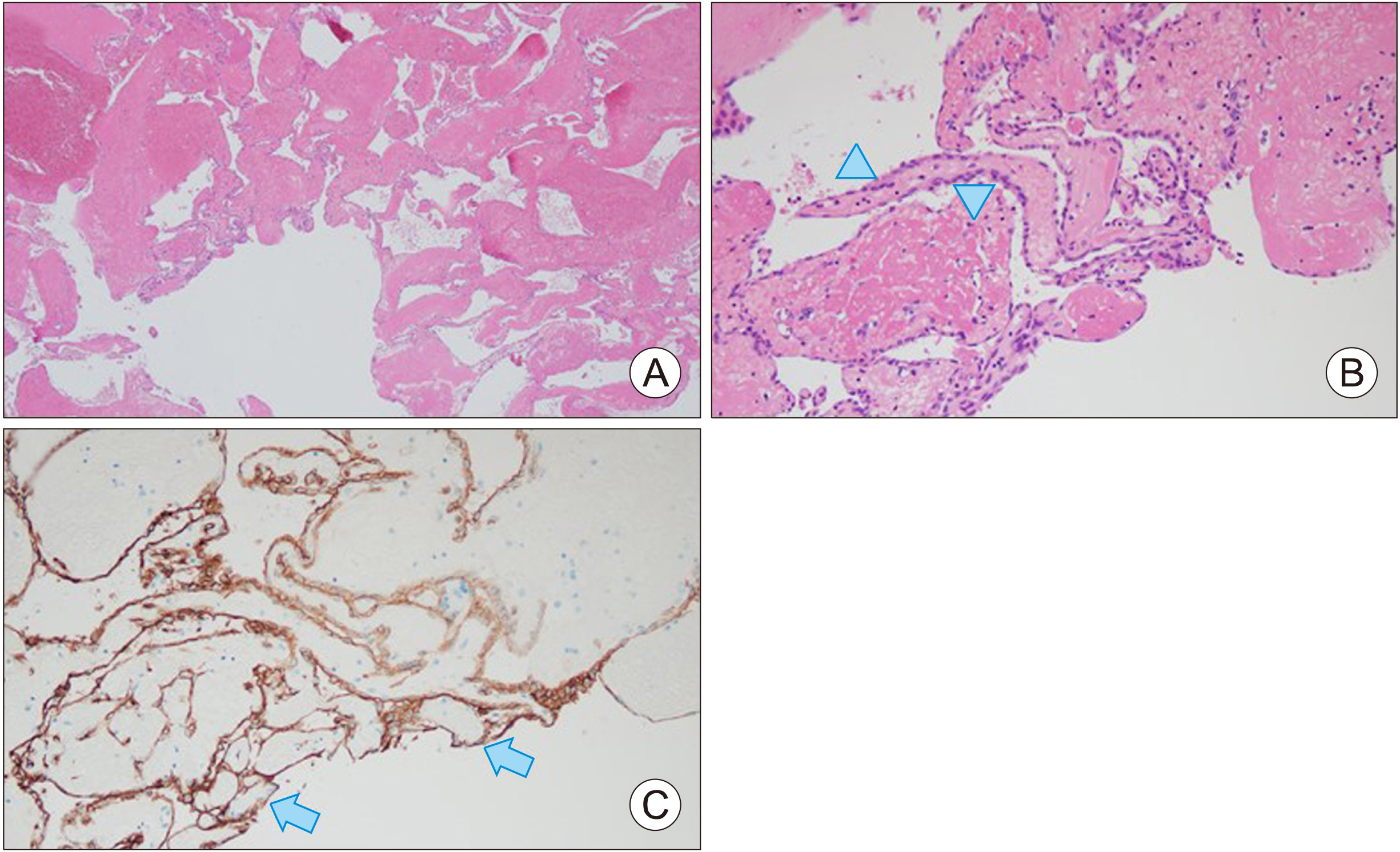
Fig. 3 Histologic slide. (A) Cavernous hemangioma composed of variably sized anastomosing vascular walls was observed at low manification (Hematoxylin and Eosin stain [H&E], ×40). (B) Each vascular wall is lined by flattened endothelial cells (H&E, ×200) (arrowheads). (C) Endothelial cells are positive for CD31 immunohistochemical stain (CD31 IHC, ×200) (arrows).
Reference
-
References
1. Masuom SHF, Amirian-Far A, Rezaei R. 2021; Primary thyroid hemangioma: a case report and literature review. Kardiochir Torakochirurgia Pol. 18(3):186–9. DOI: 10.5114/kitp.2021.109385. PMID: 34703479. PMCID: PMC8525273.2. Park SH, Kim SJ, Jung HK. 2014; Thyroid hemangiomas diagnosed on sonography. J Ultrasound Med. 33(4):729–33. DOI: 10.7863/ultra.33.4.729. PMID: 24658955.
Article3. Miao J, Chen S, Li Y, Fu L, Li H. 2017; A primary cavernous hemangioma of the thyroid gland: a case report and literature review. Medicine (Baltimore). 96(49):e8651. DOI: 10.1097/MD.0000000000008651. PMID: 29245224. PMCID: PMC5728839.4. George A, Mani V, Noufal A. 2014; Update on the classification of hemangioma. J Oral Maxillofac Pathol. 18(Suppl 1):S117–20. DOI: 10.4103/0973-029X.141321. PMID: 25364160. PMCID: PMC4211219.
Article5. Kumar B, Thangavel S, Kalaiarasi R, Ganesh RN, Saxena SK. 2020; Cavernous haemangioma of the thyroid mimicking benign nodule-a diagnostic dilemma. Int J Otorhinolaryngol Head Neck Surg. 6(12):2310–2312. DOI: 10.18203/issn.2454-5929.ijohns20205080.6. Lee J, Yun JS, Nam KH, Chung WY, Park CS. 2007; Huge cavernous hemangioma of the thyroid gland. Thyroid. 17(4):375–6. DOI: 10.1089/thy.2006.0146. PMID: 17465872.
Article7. Kumar R, Gupta R, Khullar S, Dasan B, Malhotra A. 2000; Thyroid hemangioma: a case report with a review of the literature. Clin Nucl Med. 25(10):769–71. DOI: 10.1097/00003072-200010000-00003. PMID: 11043713.8. Shpitzer T, Noyek AM, Witterick I, Kassel T, Ichise M, Gullane P, et al. 1997; Noncutaneous cavernous hemangiomas of the head and neck. Am J Otolaryngol. 18(6):367–74. DOI: 10.1016/S0196-0709(97)90055-7. PMID: 9395011.
Article9. Kano M, Kameyama K, Hosoda Y, Sugino K, Ito K. 2005; A cavernous haemangioma of the thyroid gland. J Laryngol Otol. 119(10):828–30. DOI: 10.1258/002221505774481237. PMID: 16259665.
Article10. Vanchinathan V, Mirzamani N, Kantipudi R, Schwartz EJ, Sundram UN. 2015; The vascular marker CD31 also highlights histiocytes and histiocyte-like cells within cutaneous tumors. Am J Clin Pathol. 143(2):177–85. quiz 305DOI: 10.1309/AJCPRHM8CZH5EMFD. PMID: 25596243.
