J Korean Med Sci.
2023 Nov;38(46):e399. 10.3346/jkms.2023.38.e399.
Impact of Positron Emission Tomography Viability Imaging: Guided Revascularizations on Clinical Outcomes in Patients With Myocardial Scar on Single-Photon Emission Computed Tomography Scans
- Affiliations
-
- 1Department of Internal Medicine, Kyungpook National University Hospital, Daegu, Korea
- 2School of Medicine, Kyungpook National University, Daegu, Korea
- 3Department of Nuclear Medicine, Kyungpook National University Hospital, Daegu, Korea
- KMID: 2548559
- DOI: http://doi.org/10.3346/jkms.2023.38.e399
Abstract
- Background
Positron emission tomography (PET) viability scan is used to determine whether patients with a myocardial scar on single-photon emission computed tomography (SPECT) may need revascularization. However, the clinical utility of revascularization decision-making guided by PET viability imaging has not been proven yet. The purpose of this study was to investigate the impact of PET to determine revascularization on clinical outcomes.
Methods
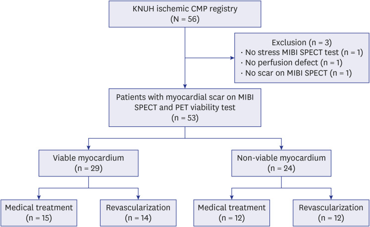
Between September 2012 and May 2021, 53 patients (37 males; mean age = 64 ± 11 years) with a myocardial scar on MIBI SPECT who underwent PET viability test were analyzed in this study. The primary outcome was a temporal change in echocardiographic findings. The secondary outcome was all-cause mortality.
Results
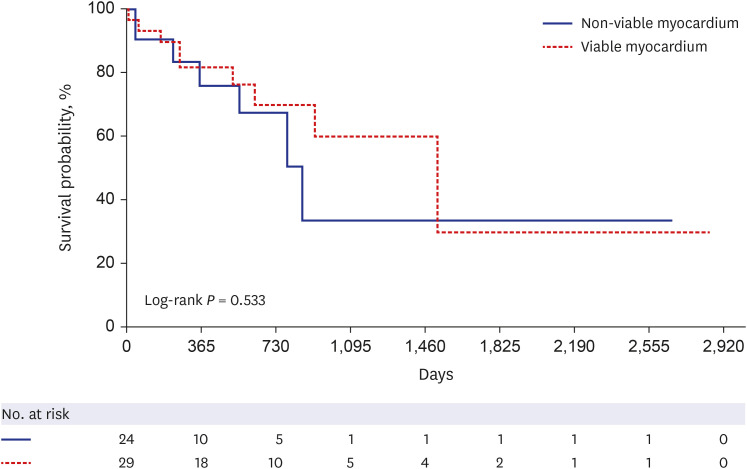
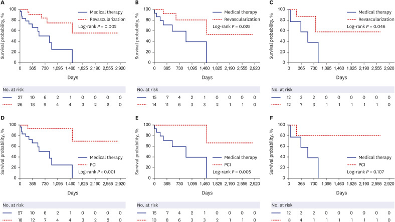
Viable myocardium was presented by PET imaging in 29 (54.7%) patients. Revascularization was performed in 26 (49.1%) patients, including 18 (34.0%) with percutaneous coronary intervention (PCI) and 8 (15.1%) with coronary artery bypass grafting. There were significant improvements in echocardiographic findings in the revascularization group and the viable myocardium group. All-cause mortality was significantly lower in the revascularization group than in the medical therapy-alone group (19.2% vs. 44.4%, log-rank P = 0.002) irrespective of viable (21.4% vs. 46.7%, log-rank P = 0.025) or non-viable myocardium (16.7% vs. 41.7%, log-rank P = 0.046). All-cause mortality was significantly lower in the PCI group than in the medical therapy-alone group (11.1% vs. 44.4%, log-rank P < 0.001).
Conclusion
Revascularization improved left ventricular systolic function and survival of patients with a myocardial scar on SPECT scans, irrespective of myocardial viability on PET scans.
Keyword
Figure
Reference
-
1. Doshi D, Ben-Yehuda O, Bonafede M, Josephy N, Karmpaliotis D, Parikh MA, et al. Underutilization of coronary artery disease testing among patients hospitalized with new-onset heart failure. J Am Coll Cardiol. 2016; 68(5):450–458. PMID: 27470451.
Article2. van Loon RB, Veen G, Baur LH, Twisk JW, van Rossum AC. Long-term follow-up of the viability guided angioplasty after acute myocardial infarction (VIAMI) trial. Int J Cardiol. 2015; 186:111–116. PMID: 25814356.
Article3. Patel H, Mazur W, Williams KA Sr, Kalra DK. Myocardial viability-State of the art: Is it still relevant and how to best assess it with imaging? Trends Cardiovasc Med. 2018; 28(1):24–37. PMID: 28735783.
Article4. Hwang HY, Yeom SY, Choi JW, Oh SJ, Park EA, Lee W, et al. Cardiac magnetic resonance predictor of ventricular function after surgical coronary revascularization. J Korean Med Sci. 2017; 32(12):2009–2015. PMID: 29115084.
Article5. Beanlands RS, Nichol G, Huszti E, Humen D, Racine N, Freeman M, et al. F-18-fluorodeoxyglucose positron emission tomography imaging-assisted management of patients with severe left ventricular dysfunction and suspected coronary disease: a randomized, controlled trial (PARR-2). J Am Coll Cardiol. 2007; 50(20):2002–2012. PMID: 17996568.
Article6. Velazquez EJ, Lee KL, Deja MA, Jain A, Sopko G, Marchenko A, et al. Coronary-artery bypass surgery in patients with left ventricular dysfunction. N Engl J Med. 2011; 364(17):1607–1616. PMID: 21463150.
Article7. Perera D, Clayton T, O’Kane PD, Greenwood JP, Weerackody R, Ryan M, et al. Percutaneous revascularization for ischemic left ventricular dysfunction. N Engl J Med. 2022; 387(15):1351–1360. PMID: 36027563.8. Felker GM, Shaw LK, O’Connor CM. A standardized definition of ischemic cardiomyopathy for use in clinical research. J Am Coll Cardiol. 2002; 39(2):210–218. PMID: 11788209.
Article9. Pellikka PA, Arruda-Olson A, Chaudhry FA, Chen MH, Marshall JE, Porter TR, et al. Guidelines for performance, interpretation, and application of stress echocardiography in ischemic heart disease: from the American Society of Echocardiography. J Am Soc Echocardiogr. 2020; 33(1):1–41.e8. PMID: 31740370.
Article10. Dilsizian V, Bacharach SL, Beanlands RS, Bergmann SR, Delbeke D, Dorbala S, et al. ASNC imaging guidelines/SNMMI procedure standard for positron emission tomography (PET) nuclear cardiology procedures. J Nucl Cardiol. 2016; 23(5):1187–1226. PMID: 27392702.
Article11. McMurray JJ, Packer M, Desai AS, Gong J, Lefkowitz MP, Rizkala AR, et al. Angiotensin-neprilysin inhibition versus enalapril in heart failure. N Engl J Med. 2014; 371(11):993–1004. PMID: 25176015.
Article12. Moss AJ, Zareba W, Hall WJ, Klein H, Wilber DJ, Cannom DS, et al. Prophylactic implantation of a defibrillator in patients with myocardial infarction and reduced ejection fraction. N Engl J Med. 2002; 346(12):877–883. PMID: 11907286.
Article13. Kim RB, Kim HS, Kang DR, Choi JY, Choi NC, Hwang S, et al. The trend in incidence and case-fatality of hospitalized acute myocardial infarction patients in Korea, 2007 to 2016. J Korean Med Sci. 2019; 34(50):e322. PMID: 31880418.
Article14. Cabac-Pogorevici I, Muk B, Rustamova Y, Kalogeropoulos A, Tzeis S, Vardas P. Ischaemic cardiomyopathy. Pathophysiological insights, diagnostic management and the roles of revascularisation and device treatment. Gaps and dilemmas in the era of advanced technology. Eur J Heart Fail. 2020; 22(5):789–799. PMID: 32020756.
Article15. Orlandini A, Castellana N, Pascual A, Botto F, Cecilia Bahit M, Chacon C, et al. Myocardial viability for decision-making concerning revascularization in patients with left ventricular dysfunction and coronary artery disease: a meta-analysis of non-randomized and randomized studies. Int J Cardiol. 2015; 182:494–499. PMID: 25617608.
Article16. Lam CS, Gamble GD, Ling LH, Sim D, Leong KT, Yeo PS, et al. Mortality associated with heart failure with preserved vs. reduced ejection fraction in a prospective international multi-ethnic cohort study. Eur Heart J. 2018; 39(20):1770–1780. PMID: 29390051.
Article
- Full Text Links
- Actions
-
Cited
- CITED
-
- Close
- Share
- Similar articles
-
- Nuclear Medicine Imaging in Rheumatic Diseases
- Recent Advances in Nuclear Cardiology
- Recent Trends in Nuclear Cardiology Practice
- 18F-2-Deoxy-2-Fluoro-D-Glucose Positron Emission Tomography: Computed Tomography for Preoperative Staging in Gastric Cancer Patients
- Practical Approach for the Clinical Use of Dopamine Transporter Imaging


