J Yeungnam Med Sci.
2023 Nov;40(Suppl):S1-S8. 10.12701/jyms.2023.00080.
Cytotoxicity of dental self-curing resin for a temporary crown: an in vitro study
- Affiliations
-
- 1Department of Dental Technology, Daegu Health College, Daegu, Korea
- 2Department of Public Health, Graduate School of Environment and Public Health Studies, Yeungnam University, Daegu, Korea
- 3Department of Occupational and Environmental Medicine, Yeungnam University Hospital, Daegu, Korea
- 4Department of Preventive Medicine and Public Health, Yeungnam University College of Medicine, Daegu, Korea
- 5Department of Dentistry, Yeungnam University College of Medicine, Daegu, Korea
- KMID: 2548336
- DOI: http://doi.org/10.12701/jyms.2023.00080
Abstract
- Background
Residual monomer tests using high-performance liquid chromatography and cytotoxicity tests were performed to analyze the effect on the oral mucosa of a self-curing resin for provisional crown production.
Methods
A cytotoxicity test was performed to confirm whether leaked residual monomers directly affected oral mucosal cells. The cytotoxicity of the liquid and solid resin polymers was measured using a water-soluble tetrazolium (WST) test and microplate reader.
Results
In the WST assay using a microplate reader, 73.4% of the cells survived at a concentration of 0.2% liquid resin polymer. The cytotoxicity of the liquid resin polymer was low at ≤0.2%. For the solid resins, when 100% of the eluate was used from each specimen, the average cell viability was 91.3% for the solid resin polymer and 100% for the hand-mixed self-curing resin, which is higher than the cell viability standard of 70%. The cytotoxicity of the solid resin polymer was low.
Conclusion
Because the polymerization process of the self-curing resin may have harmful effects on the oral mucosa during the second and third stages, the solid resin should be manufactured indirectly using a dental model.
Keyword
Figure
-

Fig. 1. The polymerization process of poly(methyl methacrylate).
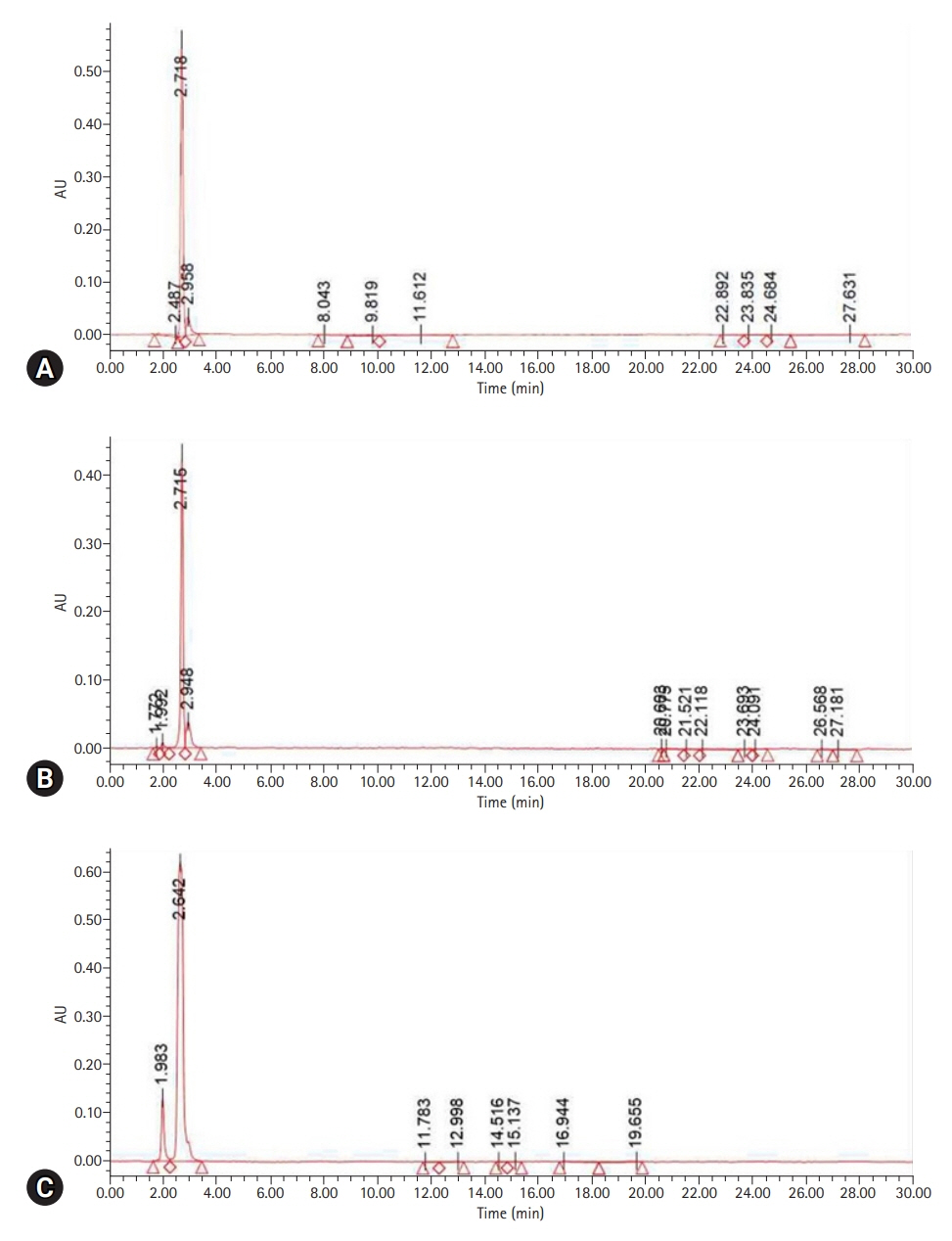
Fig. 2. Auto-scaled chromatogram of sample resin. (A) Liquid resin polymer. (B) Computer-aided design/computer-aided manufacturing self-curing resin. (C) Self-curing resin (hand-mixed). AU, absorbance unit.

Fig. 3. Effect of liquid resin polymers on cell viability.
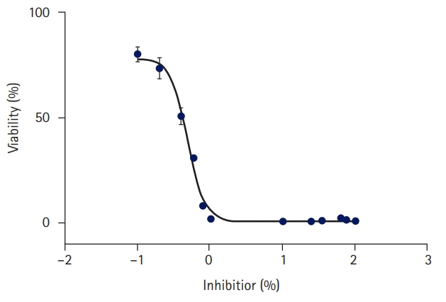
Fig. 4. Analysis of half maximal inhibitory concentration (IC50).
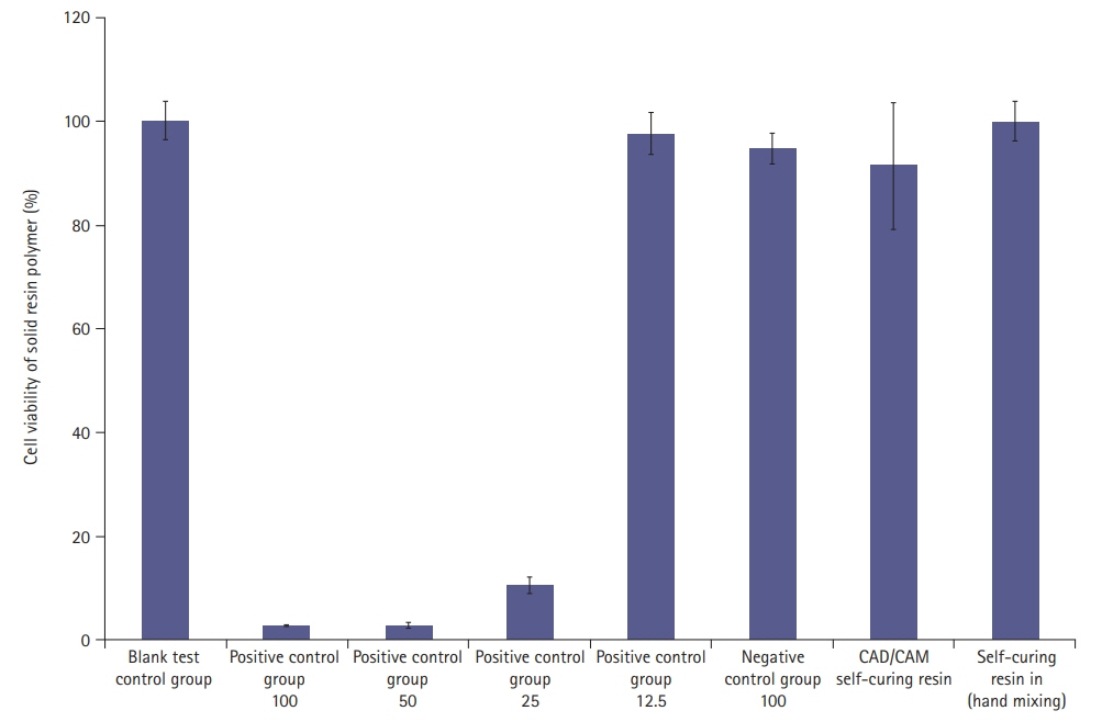
Fig. 5. Effect of solid resin polymers on cell viability. The numbers under each group name on the x-axis indicate the concentration of liquid resin polymers. CAD, computer-aided design; CAM, computer-aided manufacturing.
Reference
-
References
1. Magne P, Magne M, Belser U. The diagnostic template: a key element to the comprehensive esthetic treatment concept. Int J Periodontics Restorative Dent. 1996; 16:560–9.2. Alpert RL. A method to record optimum anterior guidance for restorative dental treatment. J Prosthet Dent. 1996; 76:546–9.
Article3. Hernandez EP, Oshida Y, Platt JA, Andres CJ, Barco MT, Brown DT. Mechanical properties of four methylmethacrylate-based resins for provisional fixed restorations. Biomed Mater Eng. 2004; 14:107–22.4. Kerby RE, Knobloch LA, Sharples S, Peregrina A. Mechanical properties of urethane and bis-acryl interim resin materials. J Prosthet Dent. 2013; 110:21–8.
Article5. Burns DR, Beck DA, Nelson SK; Committee on Research in Fixed Prosthodontics of the Academy of Fixed Prosthodontics. A review of selected dental literature on contemporary provisional fixed prosthodontic treatment: report of the Committee on Research in Fixed Prosthodontics of the Academy of Fixed Prosthodontics. J Prosthet Dent. 2003; 90:474–97.
Article6. Fondriest JF. Using provisional restorations to improve results in complex aesthetic restorative cases. Pract Proced Aesthet Dent. 2006; 18:217–23.7. Priest G. Esthetic potential of single-implant provisional restorations: selection criteria of available alternatives. J Esthet Restor Dent. 2006; 18:326–38.
Article8. Staehle HJ, Sekundo C. The origins of acrylates and adhesive technologies in dentistry. J Adhes Dent. 2021; 23:397–406.9. Harrison A, Huggett R. Effect of the curing cycle on residual monomer levels of acrylic resin denture base polymers. J Dent. 1992; 20:370–4.
Article10. Abdallah MN, Tran SD, Abughanam G, Laurenti M, Zuanazzi D, Mezour MA, et al. Biomaterial surface proteomic signature determines interaction with epithelial cells. Acta Biomater. 2017; 54:150–63.
Article11. Souto-Lopes M, Azevedo Á, Teixeira A, Bastos-Aires D, Lordelo J, Pérez-Mongiovi D. Cytotoxicity of acrylic-based resin compounds in a human gingival fibroblast cell line. Rev Port Estomatol Med Dent Cir Maxilofac. 2013; 54:87–90.
Article12. Wiegand A, Stucki L, Hoffmann R, Attin T, Stawarczyk B. Repairability of CAD/CAM high-density PMMA- and composite-based polymers. Clin Oral Investig. 2015; 19:2007–13.
Article13. Herráez-Galindo C, Rizo-Gorrita M, Luna-Oliva I, Serrera-Figallo MÁ, Castillo-Oyagüe R, Torres-Lagares D. In vitro comparative study of fibroblastic behaviour on polymethacrylate (PMMA) and lithium disilicate polymer surfaces. Polymers (Basel). 2019; 11:744.
Article14. Lee HJ, Kim CW, Kim YS. The level of residual monomer in injection molded denture base materials. J Korean Acad Prosthodont. 2003; 41:360–8.15. Kedjarune U, Charoenworaluk N, Koontongkaew S. Release of methyl methacrylate from heat-cured and autopolymerized resins: cytotoxicity testing related to residual monomer. Aust Dent J. 1999; 44:25–30.
Article16. Naji A, Harmand MF. Study of the effect of the surface state on the cytocompatibility of a Co-Cr alloy using human osteoblasts and fibroblasts. J Biomed Mater Res. 1990; 24:861–71.
Article17. Arossi GA, Lehmann M, Dihl RR, Reguly ML, de Andrade HH. Induced DNA damage by dental resin monomers in somatic cells. Basic Clin Pharmacol Toxicol. 2010; 106:124–9.
Article18. Gautam R, Singh RD, Sharma VP, Siddhartha R, Chand P, Kumar R. Biocompatibility of polymethylmethacrylate resins used in dentistry. J Biomed Mater Res B Appl Biomater. 2012; 100:1444–50.
Article19. Al-Hiyasat AS, Darmani H, Milhem MM. Cytotoxicity evaluation of dental resin composites and their flowable derivatives. Clin Oral Investig. 2005; 9:21–5.
Article20. Kang S. Mineralization-inducing potentials of calcium silicate-based pulp capping materials in human dental pulp cells. Yeungnam Univ J Med. 2020; 37:217–25.
Article21. Lai YL, Chen YT, Lee SY, Shieh TM, Hung SL. Cytotoxic effects of dental resin liquids on primary gingival fibroblasts and periodontal ligament cells in vitro. J Oral Rehabil. 2004; 31:1165–72.22. Freshney RI. Transformation and immortalization. In : Freshney RI, editor. Culture of animal cells: a manual of basic technique and specialized applications. 6th ed. New Jersey: Wiley-Blackwell;2010. p. 279–97.23. Chaves CA, Machado AL, Carlos IZ, Giampaolo ET, Pavarina AC, Vergani CE. Cytotoxicity of monomers, plasticizer and degradation by-products released from dental hard chairside reline resins. Dent Mater. 2010; 26:1017–23.
Article24. Göpferich A, Schedl L, Langer R. The precipitation of monomers during the erosion of a class of polyanhydrides. Polymer. 1996; 37:3861–9.
Article25. Jorge JH, Giampaolo ET, Machado AL, Vergani CE. Cytotoxicity of denture base acrylic resins: a literature review. J Prosthet Dent. 2003; 90:190–3.
Article26. Trivedi SC, Talim ST. The response of human gingiva to restorative materials. J Prosthet Dent. 1973; 29:73–80.
Article27. Pituru SM, Greabu M, Totan A, Imre M, Pantea M, Spinu T, et al. A review on the biocompatibility of PMMA-based dental materials for interim prosthetic restorations with a glimpse into their modern manufacturing techniques. Materials (Basel). 2020; 13:2894.
Article28. Kim EK, Park EY, Kang S. Three-dimensional printing of temporary crowns with polylactic acid polymer using the fused deposition modeling technique: a case series. J Yeungnam Med Sci. 2023; 40:302–7.
Article
- Full Text Links
-
- Actions
-
Cited
- CITED
-
- Close
- Share
-
- Similar articles
-
- Comparison of Surface Microhardness of the Flowable Bulk-Fill Resin and the Packable Bulk-Fill Resin according to Light Curing Time and Distance
- Color Stability of Self-Cured Temporary Crown Resin according to Different Surface Treatments
- A Study on the Accuracy of the record base of the Complete Denture to the Master Cast according to Kinds of Resin and Polymerization Method
- In Vitro Study on the Bond Strength Between 3D-Printed Resin and Resin Cement for Pediatric Crown Restoration
- Comparison of polymerization shrinkage of dual-cure core build-up resin according to shade and curing mode
