Merging CT on Fluoroscopic Imaging Using the Trachea as a Reference Without Contrast
- Affiliations
-
- 1Division of Cardiology, Department of Internal Medicine, Keimyung University Dongsan Hospital, Daegu, Korea
- 2Department of Radiology, Keimyung University Dongsan Hospital, Daegu, Korea
- KMID: 2548242
- DOI: http://doi.org/10.4070/kcj.2023.0190
Figure
-
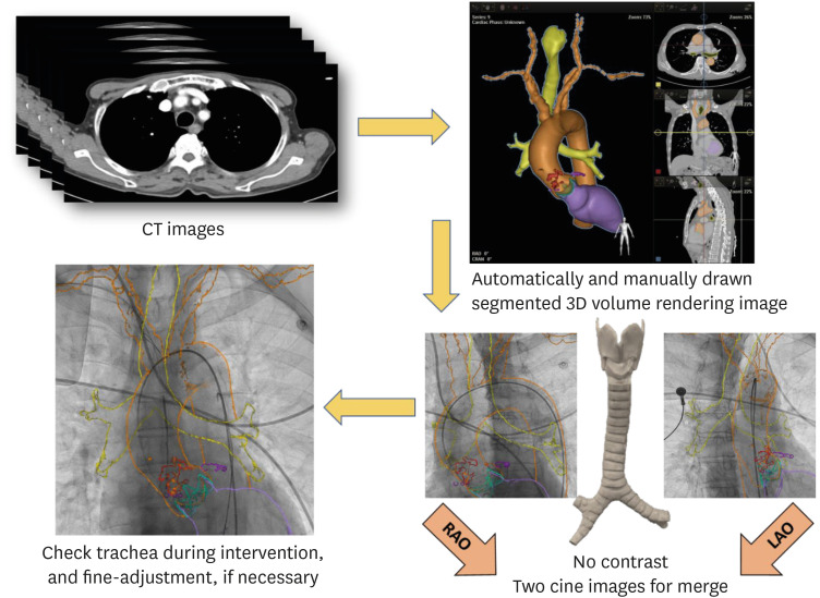
Figure 1 Procedural process of computed tomography merged imaging with the trachea as reference.After uploading the raw CT images to the HeartNavigator - Structural Heart Disease system, a segmented volume rendering image is created. The RAO and LAO view cine images are used to focus the patient and CT images for the procedure. If minor deviations are observed, fine adjustment can be considered perioperatively.CT = computed tomography; LAO = left anterior oblique; RAO = right anterior oblique.
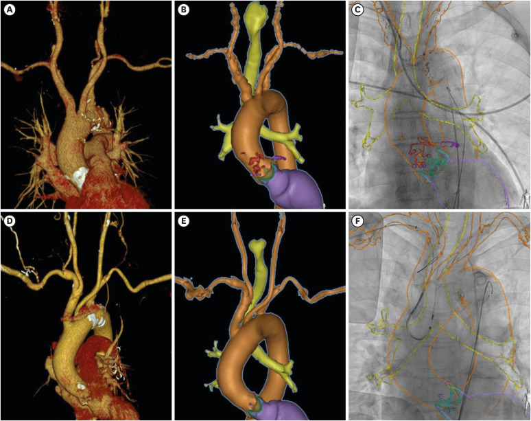
Figure 2 Images of CT merged with cerebral protection system insertion.Conventional volume rendering images of cerebral protection system candidate patient (case 1) (A), automatically and manually drawn segmented volume rendering image by HeartNavigator - Structural Heart Disease system (B), procedural CT merged image (C), CT images of case 2 (D, E, and F).CT = computed tomography.
