Restor Dent Endod.
2023 Aug;48(3):e22. 10.5395/rde.2023.48.e22.
Tip and taper compatibility of accessory gutta-percha points with rotary and reciprocating instruments
- Zanatta Streck JN
 1
1 - Arcaro S1
- Ceretta RA1
- Bortoluzzi EA2
- Roberti Garcia LdF3
- Almeida Jd4
- Poli Kopper PM5
- Bernardi AV1,6
- Affiliations
-
- 1Department of Endodontics, School of Dentistry, University of Extreme Southern Santa Catarina, Criciúma, SC, Brazil
- 2Department of Diagnosis & Oral Health, Division of Endodontics, School of Dentistry, University of Louisville, Louisville, KY, USA
- 3Department of Dentistry - Endodontics Division, Federal University of Santa Catarina, Florianopolis, SC, Brazil
- 4Department of Endodontics, School of Dentistry, University of Southern Santa Catarina, Florianópolis, SC, Brazil
- 5Program in Dentistry, School of Dentistry, Federal University of Rio Grande do Sul, Porto Alegre, RS, Brazil
- 6Biomaterials Group, Graduate Program in Materials Science and Engineering, University of Extreme Southern Santa Catarina, Criciúma, SC, Brazil
- KMID: 2548225
- DOI: http://doi.org/10.5395/rde.2023.48.e22
Abstract
Objectives
This study was conducted to evaluate and compare the tip and taper compatibility of accessory gutta-percha points (AGPs) with various rotary and reciprocating instruments.
Materials and Methods
Using a profile analyzer, tip and taper measurements were taken of 10 AGPs of each of the 14 models available from Odous de Deus and the 4 models available from Dentsply-Maillefer. Diameter measurements were taken at 1-mm intervals, from 3 mm from the tip (D3) to 16 mm.
Results
Based on the mean values obtained, 3-dimensional (3D) models of the AGPs were drawn in Autodesk Fusion 360 and superimposed on 3D models of each instrument selected (Mtwo, Reciproc, RaCe, K3, and ProDesign Logic) to determine the compatibility between the instrument and the AGP. Data corresponding to the tips and tapers of the various AGPs, as well as the tip and taper differences between the AGPs and the instruments, were analyzed using descriptive statistics. The tapers of the AGPs were subject to the American National Standards Institute/American Dental Association No. 57 standard. The Odous de Deus extralong medium and extra-long extra-medium AGPs were shown to be compatible with Mtwo, K3, and ProDesign Logic instruments with taper 0.06 and tip sizes 25 and 30, while the Dentsply fine and fine medium cones were compatible with Mtwo, RaCe, and K3 instruments with conicity of 0.04 and tip sizes 35 and 40.
Conclusions
Both the Odous de Deus and Dentsply commercial brands included 2 AGP models with tip (D3) and taper compatibility with Mtwo, RaCe, K3, and/or Prodesign Logic instruments.
Keyword
Figure
-
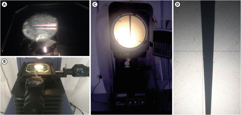
Figure 1 Measurement of accessory gutta-percha points.(A) Cone positioned on the projector table. (B) Projector table and X and Y digital counters. (C) Profile analyzer model PJ-A3000, Mitutoyo American Corporation. (D) Cone projection screen with X and Y axes.
Reference
-
1. Ng YL, Mann V, Rahbaran S, Lewsey J, Gulabivala K. Outcome of primary root canal treatment: systematic review of the literature -- Part 2. Influence of clinical factors. Int Endod J. 2008; 41:6–31. PMID: 17931388.
Article2. Pedro FM, Marques A, Pereira TM, Bandeca MC, Lima S, Kuga MC, Tonetto MR, Semenoff-Segundo A, Borges AH. Status of endodontic treatment and the correlations to the quality of root canal filling and coronal restoration. J Contemp Dent Pract. 2016; 17:830–836. PMID: 27794154.
Article3. Tomson RM, Polycarpou N, Tomson PL. Contemporary obturation of the root canal system. Br Dent J. 2014; 216:315–322. PMID: 24651337.
Article4. Vishwanath V, Rao HM. Gutta-percha in endodontics - a comprehensive review of material science. J Conserv Dent. 2019; 22:216–222. PMID: 31367101.
Article5. Ricucci D, Rôças IN, Alves FR, Loghin S, Siqueira JF Jr. Apically extruded sealers: fate and influence on treatment outcome. J Endod. 2016; 42:243–249. PMID: 26725179.
Article6. Patni PM, Chandak M, Jain P, Patni MJ, Jain S, Mishra P, Jain V. Stereomicroscopic evaluation of sealing ability of four different root canal sealers- an in vitro study. J Clin Diagn Res. 2016; 10:ZC37–ZC39.7. Marashdeh MQ, Friedman S, Lévesque C, Finer Y. Esterases affect the physical properties of materials used to seal the endodontic space. Dent Mater. 2019; 35:1065–1072. PMID: 31104923.
Article8. Ørstavik D, Nordahl I, Tibballs JE. Dimensional change following setting of root canal sealer materials. Dent Mater. 2001; 17:512–519. PMID: 11567689.
Article9. Schäfer E, Köster M, Bürklein S. Percentage of gutta-percha-filled areas in canals instrumented with nickel-titanium systems and obturated with matching single cones. J Endod. 2013; 39:924–928. PMID: 23791265.
Article10. Yürüker S, Görduysus M, Küçükkaya S, Uzunoğlu E, Ilgın C, Gülen O, Tuncel B, Görduysus MÖ. Efficacy of combined use of different nickel-titanium files on removing root canal filling materials. J Endod. 2016; 42:487–492. PMID: 26778268.
Article11. Coelho MS, Card SJ, Tawil PZ. Safety assessment of two hybrid instrumentation techniques in a dental student endodontic clinic: a retrospective study. J Dent Educ. 2017; 81:333–339. PMID: 28250040.
Article12. Council on Dental Materials, Instruments, and Equipment. ANSI/ADA specification no. 57 for endodontic filling materials. updated November 28, 2022. cited September 15, 2022. Available from: . DOI: 10.14219/jada.archive.1984.0208.13. Haupt F, Seidel M, Rizk M, Sydow HG, Wiegand A, Rödig T. Diameter and taper variability of single-file instrumentation systems and their corresponding gutta-percha cones. J Endod. 2018; 44:1436–1441. PMID: 30078573.
Article14. Lask JT, Walker MP, Kulild JC, Cunningham KP, Shull PA. Variability of the diameter and taper of size #30, 0.04 nickel-titanium rotary files. J Endod. 2006; 32:1171–1173. PMID: 17174675.
Article15. Holland R, Gomes JE, Cintra LT, Queiroz ÍO, Estrela C. Factors affecting the periapical healing process of endodontically treated teeth. J Appl Oral Sci. 2017; 25:465–476. PMID: 29069143.
Article16. Ersahan S, Aydin C. Solubility and apical sealing characteristics of a new calcium silicate-based root canal sealer in comparison to calcium hydroxide-, methacrylate resin- and epoxy resin-based sealers. Acta Odontol Scand. 2013; 71:857–862. PMID: 23088627.
Article17. Rosen E, Goldberger T, Taschieri S, Del Fabbro M, Corbella S, Tsesis I. The prognosis of altered sensation after extrusion of root canal filling materials: a systematic review of the literature. J Endod. 2016; 42:873–879. PMID: 27133502.
Article18. Ricucci D, Lin LM, Spångberg LS. Wound healing of apical tissues after root canal therapy: a long-term clinical, radiographic, and histopathologic observation study. Oral Surg Oral Med Oral Pathol Oral Radiol Endod. 2009; 108:609–621. PMID: 19716731.
Article19. Gordon MP, Love RM, Chandler NP. An evaluation of .06 tapered gutta-percha cones for filling of .06 taper prepared curved root canals. Int Endod J. 2005; 38:87–96. PMID: 15667630.
Article20. Almeida BM, Provenzano JC, Marceliano-Alves MF, Rôças IN, Siqueira JF Jr. Matching the dimensions of currently available instruments with the apical diameters of mandibular molar mesial root canals obtained by micro-computed tomography. J Endod. 2019; 45:756–760. PMID: 31056298.
Article21. Brunson M, Heilborn C, Johnson DJ, Cohenca N. Effect of apical preparation size and preparation taper on irrigant volume delivered by using negative pressure irrigation system. J Endod. 2010; 36:721–724. PMID: 20307751.
Article22. Keles A, Keskin C, Alqawasmi R, Versiani MA. Evaluation of dentine thickness of middle mesial canals of mandibular molars prepared with rotary instruments: a micro-CT study. Int Endod J. 2020; 53:519–528. PMID: 31705697.
Article23. Metzger Z, Nissan R, Tagger M, Tamse A. Apical seal by customized versus standardized master cones: a comparative study in flat and round canals. J Endod. 1988; 14:381–384. PMID: 3253401.
Article24. Silvestrin T, Torabinejad M, Handysides R, Shabahang S. Effect of apex size on the leakage of gutta-percha and sealer-filled root canals. Quintessence Int. 2016; 47:373–378. PMID: 26824086.25. American National Standard/American Dental Association Standard (ANSI/ADA). Specification No. 58. Root canal files, type H (Hedstrom). updated November 28, 2022. cited September 15th, 2022. Available from: https://webstore.ansi.org/preview-pages/ADA/preview_ANSI+ADA+58-2010+R2015.pdf.
- Full Text Links
-
- Actions
-
Cited
- CITED
-
- Close
- Share
-
- Similar articles
-
- Obturation efficiency of non-standardized gutta-percha cone in curved root canals prepared with 0.06 taper nickel-titanium instruments
- A study of insertion depth of gutta percha cones after shaping by Ni-Ti rotary files in simulated canals
- The effect of gutta-percha removal using nickel-titanium rotary instruments
- A comparison of thermoplasticized injectable gutta-percha techniques in ribbon-shaped canals : adaptation to canal walls
- Comparison of warm gutta-percha condensation techniques in ribbon shaped canal: weight of filled gutta-percha
