Fibromuscular Dysplasia Involving the Iliac Artery and Mimicking Atresia of the Left Iliac Artery
- Affiliations
-
- 1Division of Cardiology, Department of Internal Medicine, Ewha Womans University Mokdong Hospital, Ewha Womans University College of Medicine, Seoul, Korea
- KMID: 2547925
- DOI: http://doi.org/10.12771/emj.2023.e21
Figure
-

Fig. 1. CT angiography findings of the lower extremities. (A) The left iliac artery is completely occluded from the aortic bifurcation (circle; “string of beads” appearance of the right external iliac artery from invasive angiography). (B) Atherosclerotic plaques are present in the abdominal aorta and (C) at the bifurcation site. (D) A thread-like hypoplastic left iliac artery is visible. (E−G) Multiple layers of thread-like stenosis (red arrows) and a beaded appearance of the left popliteal artery are evident on CT angiography (orange arrows).
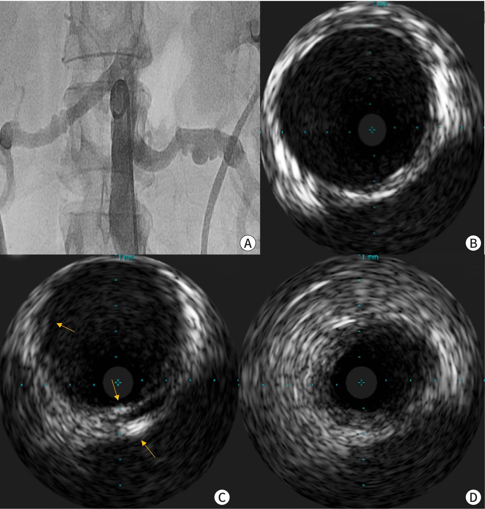
Fig. 2. Invasive images of the renal arteries. (A) Angiography reveals the typical “string of beads” appearance in both renal arteries. Intravascular ultrasonography indicates varied disease progression. (B) The vessel layers are relatively preserved. (C) There is an intimal fibromuscular ridge and increased medial echogenicity, as indicated by the arrows. (D) Circumferential hyperplasia of the intimal or medial layers is noted, accompanied by denudation of the vessel layers.
Reference
-
References
1. Olin JW, Gornik HL, Bacharach JM, Biller J, Fine LJ, Gray BH, et al. Fibromuscular dysplasia: state of the science and critical unanswered questions: a scientific statement from the American Heart Association. Circulation. 2014; 129((9)):1048–1078. DOI: 10.1161/01.cir.0000442577.96802.8c. PMID: 24548843.
- Full Text Links
-
- Actions
-
Cited
- CITED
-
- Close
- Share
-
- Similar articles
-
- Treatment of Fibromuscular Dysplasia of the Abdominal Aorta Causing Renovascular Hypertension and Aneurysm: A case report
- Hypoplasia of Left Vertebral Artery with Intimal Fibromuscular Dysplasia in a Korean Woman
- Ureteral Stricture from Retroperitoneal Fibrosis Caused by Isolated Common Iliac Artery Aneurysm
- Fibromuscular Dysplasia of the Distal Internal Carotid and Middle Cerebral Artery
- The inferior epigastric artery arising from the internal iliac artery via a common trunk with the obturator artery
