Evaluation of the mechanical properties of current biliary self-expandable metallic stents: axial and radial force, and axial force zero border
- Yamagata W
 1
1 - Fujisawa T1
- Sasaki T2
- Ishibashi R3
- Saito T3
- Yoshida S4
- No S5
- Inoue K5
- Nakai Y3,4
- Sasahira N2
- Isayama H1
- Affiliations
-
- 1Department of Gastroenterology, Graduate School of Medicine, Juntendo University, Tokyo, Japan
- 2Department of Hepato-Biliary-Pancreatic Medicine, Cancer Institute Hospital of Japanese Foundation for Cancer Research, Tokyo, Japan
- 3Department of Gastroenterology, Graduate School of Medicine, The University of Tokyo, Tokyo, Japan
- 4Department of Endoscopy and Endoscopic Surgery, Graduate School of Medicine, The University of Tokyo, Tokyo, Japan
- 5Medical Laboratory, Research & Development Center, Zeon Corporation, Toyama, Japan
- KMID: 2546142
- DOI: http://doi.org/10.5946/ce.2022.201
Abstract
- Background/Aims
Mechanical properties (MPs) and axial and radial force (AF and RF) may influence the efficacy and complications of self-expandable metallic stent (SEMS) placement. We measured the MPs of various SEMSs and examined their influence on the SEMS clinical ability.
Methods
We evaluated the MPs of 29 types of 10-mm SEMSs. RF was measured using a conventional measurement device. AF was measured using the conventional and new methods, and the correlation between the methods was evaluated.
Results
A high correlation in AFs was observed, as measured by the new and conventional manual methods. AF and RF scatterplots divided the SEMSs into three subgroups according to structure: hook-and-cross-type (low AF and RF), cross-type (high AF and low RF), and laser-cut-type (intermediate AF and high RF). The hook-and-cross-type had the largest axial force zero border (>20°), followed by the laser-cut and cross types.
Conclusions
MPs were related to stent structure. Hook-and-cross-type SEMSs had a low AF and high axial force zero border and were considered safest because they caused minimal stress on the biliary wall. However, the increase in RF must be overcome.
Keyword
Figure
-

Fig. 1. Types of biliary self-expandable metallic stents. (A) Cross-type stents are knitted to form an X-shape, and the wires do not separate (WallFlex). (B) Hook-and-cross-type stents are formed by hook and cross knitting, in varying proportions. In hook knitting, the wires form a V-shape, and are separated from each other by bends (Hanaro). (C) Zigzag-type stents consist of a wavy wire shaped into vertically connected rings forming a cylinder. The laser-cut stent is based on the zigzag stent (EPIC).

Fig. 2. Overview of the self-expandable metallic stents evaluated in the present study.

Fig. 3. Radial force measurement device. The stent samples were inserted into the cylinder and then expanded. Expansion and resistance forces were measured.

Fig. 4. Relationship between hoop force (HF) and radial force. The HF was modified to accord with the original concept of radial force. Radial force was calculated by multiplying HF by 2π (6.28). RF, radial force.

Fig. 5. (A) Conventional manual method of measuring axial force (AF). This method measures the linearizing force of self-expandable metallic stent bending at 60°. (B) New AF measurement method. This method automatically measures torque in a straight stent position using an AF measurement device.

Fig. 6. Axial force (AF) measurement method using the new device. (A) AF measurement at onset. (B) AF measurement during the bending phase. The yellow arrow in the photo indicates the direction in which the arm moves. (C) Transition from the bending to straightening phase at 90°. (D) AF measurement during the straightening phase. The axial force zero border was measured as the angle at which the torque force was <0.05 mNm during the straightening phase.
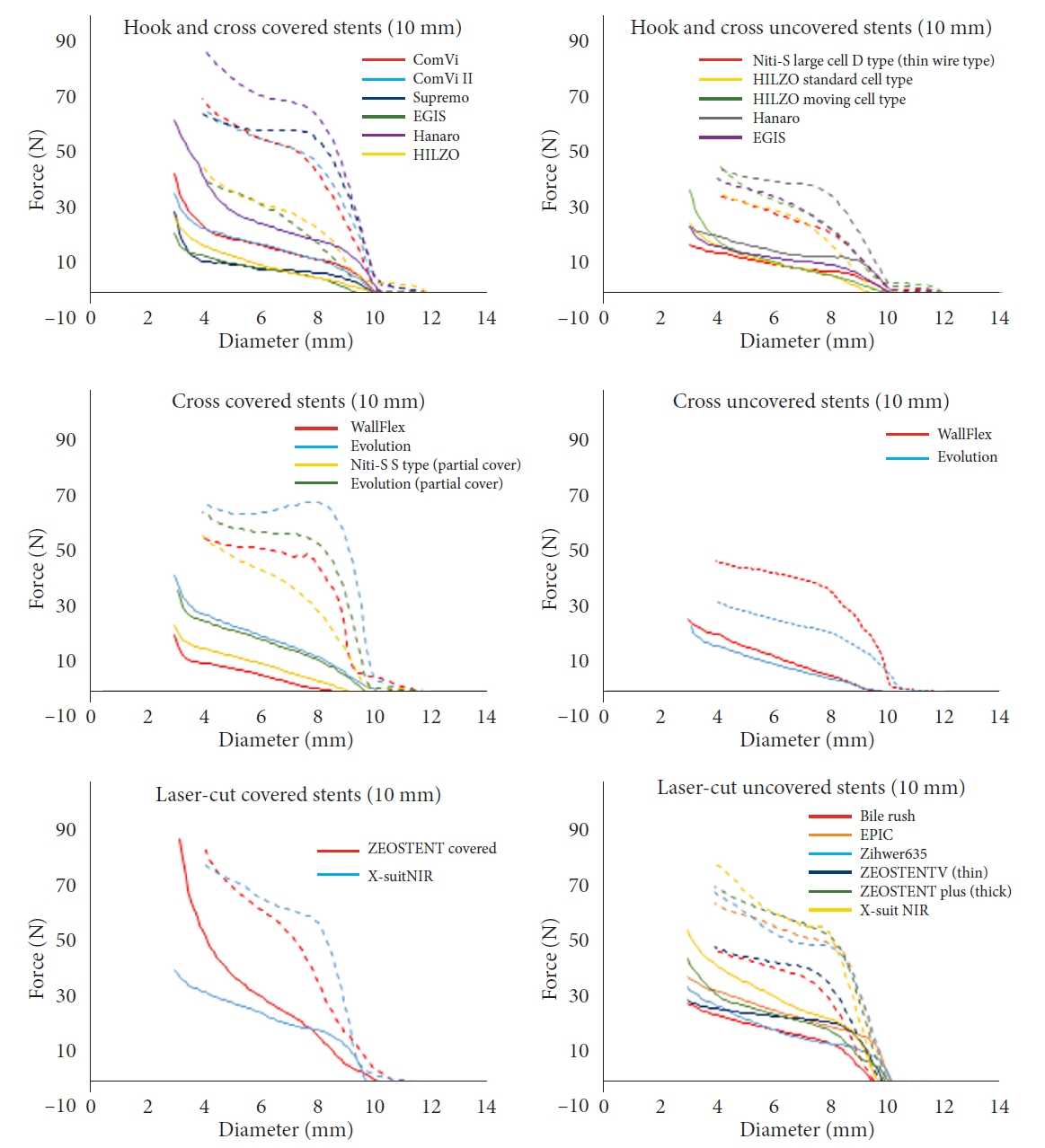
Fig. 7. Radial force versus stent diameter curves. The same self-expandable metallic stents type, differing only in diameter, had similar curves. Expansion force was first measured using the cylinder until the stent was fully expanded. The diameter of the cylinder decreased, and the resistance force was measured (solid line: expansion force; dotted line: resistance force).
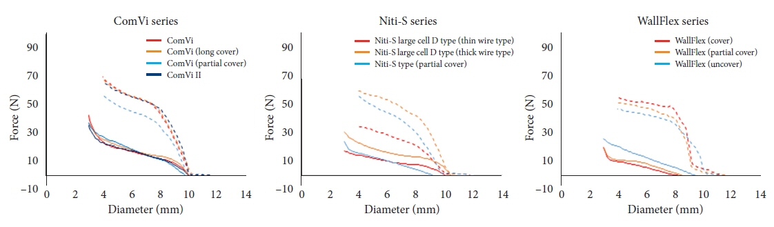
Fig. 8. Radial force versus stent diameter curve for the ComVi, WallFlex, and Niti-S stents (solid line: expansion force; dotted line: resistance force).
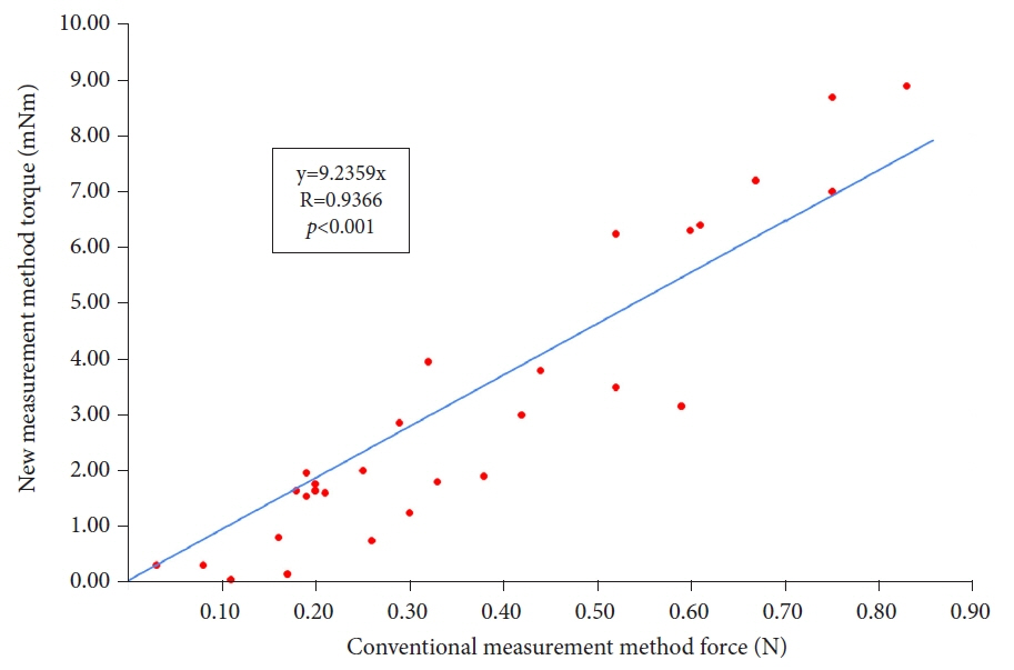
Fig. 9. Axial force measured by the new and conventional methods; a strong correlation was found (y=9.2359x, R=0.9366, p<0.001).
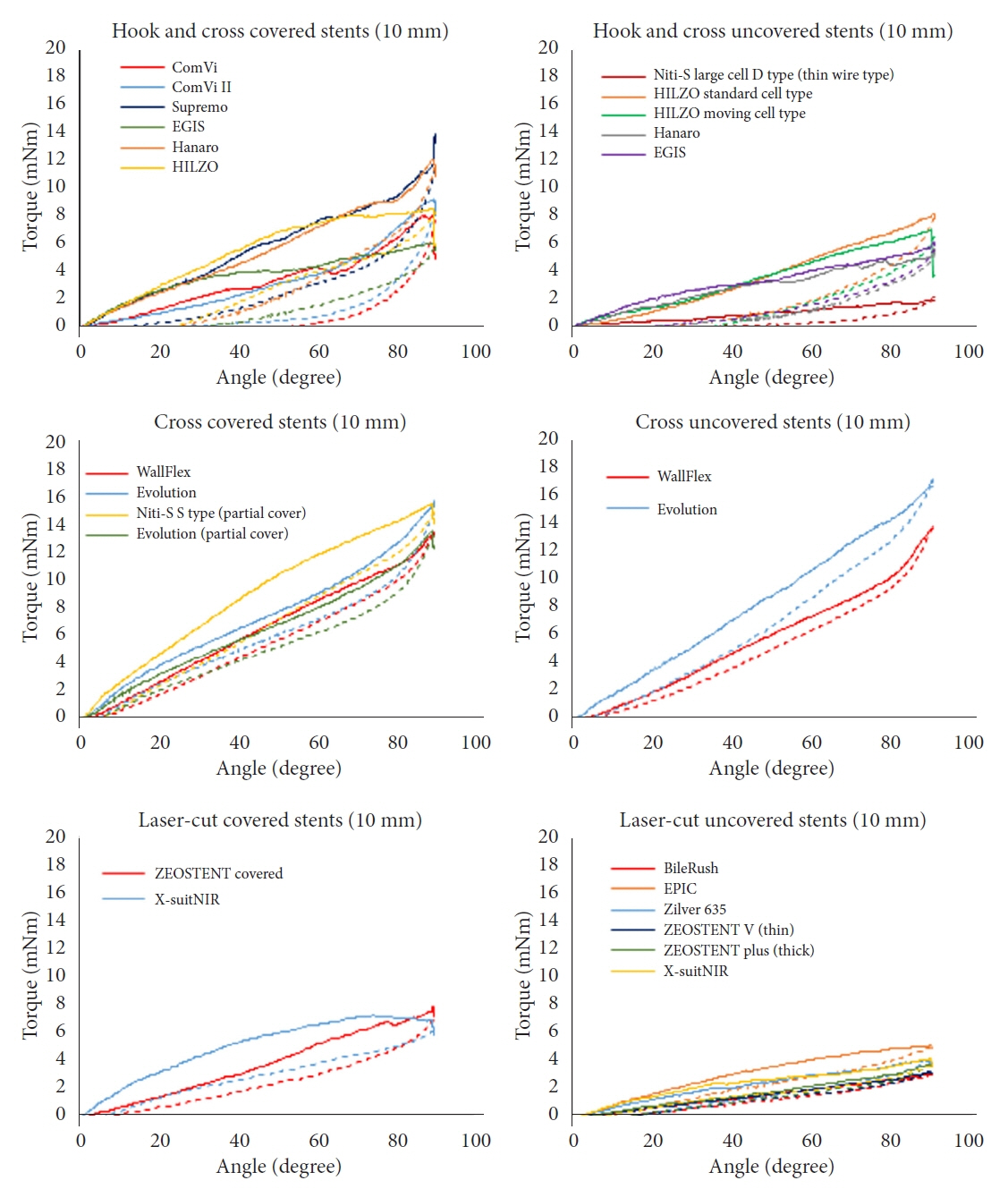
Fig. 10. Axial force versus angle curves. The axial force of stents of various diameters was measured using the new measurement device. The stent was bent to 90° and then straightened (solid line: bending phase; dotted line: straightening phase).
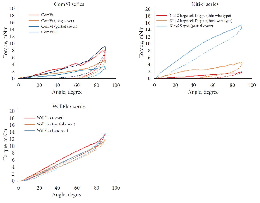
Fig. 11. Axial force versus stent angle curves for ComVi, WallFlex, and Niti-S series stents (solid line: bending phase; dotted line: straightening phase).

Fig. 12. Axial force zero border (red, hook-and-cross type; yellow, cross-type; blue, laser-cut-type).
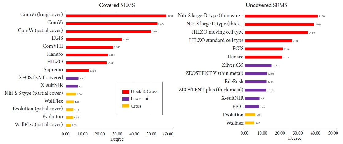
Fig. 13. Radial (RF) and axial force (AF) scatterplots of 10 mm self-expandable metallic stent (SEMS). Outward RF at 4 mm and AF in the straightening phase with the stent at 60°, as measured by the new method, are plotted (red, hook-and-cross type; yellow, cross-type; blue, laser-cut-type).

Cited by 1 articles
-
How to reduce fistula formation after self-expandable metallic stent insertion for treating malignant esophageal stricture?
Kwang Bum Cho
Clin Endosc. 2023;56(6):735-737. doi: 10.5946/ce.2023.257.
Reference
-
1. Conio M, Mangiavillano B, Caruso A, et al. Covered versus uncovered self-expandable metal stent for palliation of primary malignant extrahepatic biliary strictures: a randomized multicenter study. Gastrointest Endosc. 2018; 88:283–291.2. Sawas T, Al Halabi S, Parsi MA, et al. Self-expandable metal stents versus plastic stents for malignant biliary obstruction: a meta-analysis. Gastrointest Endosc. 2015; 82:256–267.3. Lam R, Muniraj T. Fully covered metal biliary stents: a review of the literature. World J Gastroenterol. 2021; 27:6357–6373.4. Kitagawa K, Mitoro A, Ozutsumi T, et al. Laser-cut-type versus braided-type covered self-expandable metallic stents for distal biliary obstruction caused by pancreatic carcinoma: a retrospective comparative cohort study. Clin Endosc. 2022; 55:434–442.5. Isayama H, Mukai T, Itoi T, et al. Comparison of partially covered nitinol stents with partially covered stainless stents as a historical control in a multicenter study of distal malignant biliary obstruction: the WATCH study. Gastrointest Endosc. 2012; 76:84–92.6. Saito H, Sakurai Y, Takamura A, et al. [Biliary endoprosthesis using Gore-Tex covered expandable metallic stents: preliminary clinical evaluation]. Nihon Igaku Hoshasen Gakkai Zasshi. 1994; 54:180–182.7. Hori Y, Hayashi K, Yoshida M, et al. Novel characteristics of traction force in biliary self-expandable metallic stents. Dig Endosc. 2017; 29:347–352.8. U.S. Department of Health and Human Services, Food and Drug Administration. Center for Devices and Radiological Health. Metal expandable biliary stents: premarket notification (510(k)) submissions. Silver Spring: Center for Drug Evaluation and Research;2019.9. Isayama H, Nakai Y, Toyokawa Y, et al. Measurement of radial and axial forces of biliary self-expandable metallic stents. Gastrointest Endosc. 2009; 70:37–44.10. Katsinelos P, Lazaraki G, Gkagkalis S, et al. A fully covered self-expandable metal stent anchored by a 10-Fr double pigtail plastic stent: an effective anti-migration technique. Ann Gastroenterol. 2017; 30:114–117.11. Isayama H, Nakai Y, Hamada T, et al. Understanding the mechanical forces of self-expandable metal stents in the biliary ducts. Curr Gastroenterol Rep. 2016; 18:64.12. Nakai Y, Isayama H, Kawakubo K, et al. Metallic stent with high axial force as a risk factor for cholecystitis in distal malignant biliary obstruction. J Gastroenterol Hepatol. 2014; 29:1557–1562.13. Jang S, Stevens T, Parsi M, et al. Association of covered metallic stents with cholecystitis and stent migration in malignant biliary stricture. Gastrointest Endosc. 2018; 87:1061–1070.14. Kawakubo K, Isayama H, Nakai Y, et al. Risk factors for pancreatitis following transpapillary self-expandable metal stent placement. Surg Endosc. 2012; 26:771–776.15. Takeda T, Sasaki T, Mie T, et al. Novel risk factors for recurrent biliary obstruction and pancreatitis after metallic stent placement in pancreatic cancer. Endosc Int Open. 2020; 8:E1603–E1610.16. Nakai Y, Isayama H, Kogure H, et al. Risk factors for covered metallic stent migration in patients with distal malignant biliary obstruction due to pancreatic cancer. J Gastroenterol Hepatol. 2014; 29:1744–1749.17. Sasaki T, Ishibashi R, Yoshida S, et al. Comparing the mechanical properties of a self-expandable metallic stent for colorectal obstruction: proposed measurement method of axial force using a new measurement machine. Dig Endosc. 2021; 33:170–178.18. Rodrigues-Pinto E, Morais R, Sousa-Pinto B, et al. Development of an online app to predict post-endoscopic retrograde cholangiopancreatography adverse events using a single-center retrospective cohort. Dig Dis. 2021; 39:283–293.19. Ida Bagus B. A rare clinical presentation of third part duodenal perforation due to post-endoscopic retrograde cholangiopancreatography stent migration on advanced stage peri-ampullary tumor. JGH Open. 2021; 5:968–970.20. Mukai T, Yasuda I, Isayama H, et al. Comparison of axial force and cell width of self-expandable metallic stents: which type of stent is better suited for hilar biliary strictures? J Hepatobiliary Pancreat Sci. 2011; 18:646–652.21. Weaver M, Lang G, Das K, et al. Metal biliary stent erosion through the common bile duct leading to obstructive jaundice and cholangitis. Am J Gastroenterol. 2021; 116:1372.
- Full Text Links
-
- Actions
-
Cited
- CITED
-
- Close
- Share
-
- Similar articles
-
- Mechanical Property and Problems of the Self-expandable Metal Stent in Pancreaticobiliary Cancer
- Mechanical Characteristics of Self-expandable Metallic Stents: In Vitro Study with Three of Stress
- Metallic EndoCoilTM Stent Application for Patients with Malignant Obstructive Jaundice
- Basic Knowledge about Metal Stent Development
- Mechanical Characterization of Self-Expandable Esophageal Metal Stents
