J Korean Assoc Oral Maxillofac Surg.
2023 Aug;49(4):214-217. 10.5125/jkaoms.2023.49.4.214.
Case series and technical report of nasal floor approach for mesiodens
- Affiliations
-
- 1Department of Oral and Maxillofacial Surgery, School of Dentistry and Institute of Oral Bioscience, Research Institute of Clinical Medicine of Jeonbuk National University-Biomedical Research Institute of Jeonbuk National University Hospital, Jeonbuk National University, Jeonju, Korea
- KMID: 2545587
- DOI: http://doi.org/10.5125/jkaoms.2023.49.4.214
Abstract
Objectives
This case series aims to introduce the nasal floor approach for extracting inverted mesiodens.
Materials and Methods
Through a retrospective chart review between January 2022 and February 2023, we included the mesiodens patients using nasal floor approach, and analysis the location of mesiodens from the anterior nasal spine (ANS), total operation time, and complications.
Results
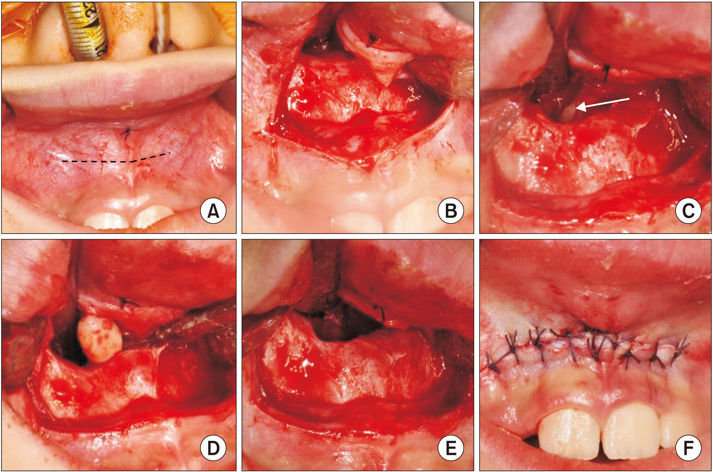
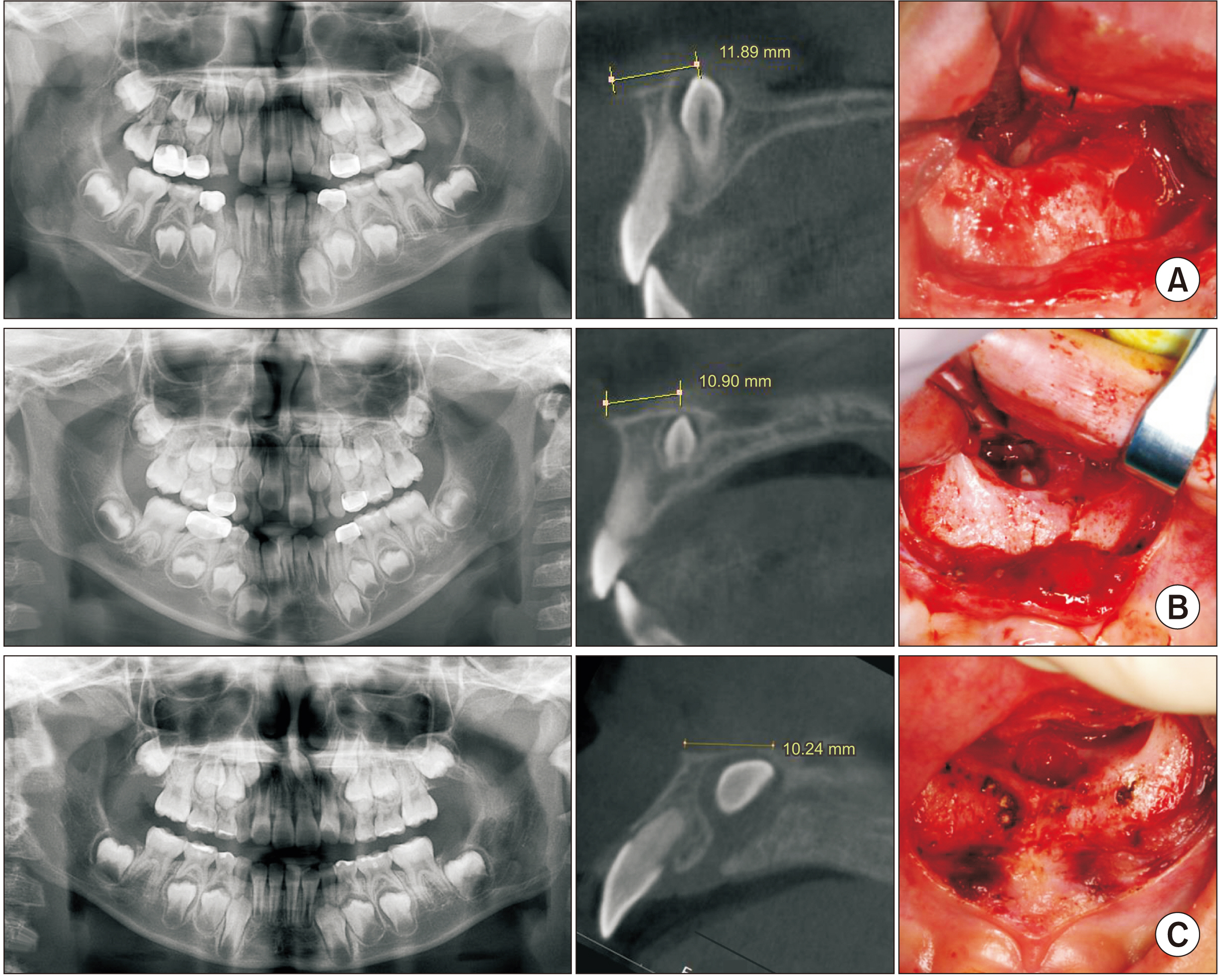
Each mesiodens was located 10 to 12 mm from the ANS and was covered with a cortical layer of the nasal floor. All mesiodens were successfully extracted without exposing the adjacent incisors or nasopalatine nerve within 30 minutes from draping to postoperative dressing.
Conclusion
The nasal floor approach is an efficient extraction method that reduces bone removal and prevents anatomical damage while removing the mesiodens just below the nasal floor bone.
Figure
Reference
-
References
1. Syriac G, Joseph E, Rupesh S, Philip J, Cherian SA, Mathew J. 2017; Prevalence, characteristics, and complications of supernumerary teeth in nonsyndromic pediatric population of South India: a clinical and radiographic study. J Pharm Bioallied Sci. 9(Suppl 1):S231–6. https://doi.org/10.4103/jpbs.jpbs_154_17. DOI: 10.4103/jpbs.JPBS_154_17. PMID: 29284970. PMCID: PMC5731020.
Article2. Shih WY, Hsieh CY, Tsai TP. 2016; Clinical evaluation of the timing of mesiodens removal. J Chin Med Assoc. 79:345–50. https://doi.org/10.1016/j.jcma.2015.10.013. DOI: 10.1016/j.jcma.2015.10.013. PMID: 27090104.
Article3. Mukhopadhyay S. 2011; Mesiodens: a clinical and radiographic study in children. J Indian Soc Pedod Prev Dent. 29:34–8. https://doi.org/10.4103/0970-4388.79928. DOI: 10.4103/0970-4388.79928. PMID: 21521916.
Article4. Mathew M, Sasikumar P, Vivek VJ, Kala R. 2020; Surgical management of a rare case of inverted intranasal mesiodens. BMJ Case Rep. 13:e237982. https://doi.org/10.1136/bcr-2020-237982. DOI: 10.1136/bcr-2020-237982. PMID: 33127709. PMCID: PMC7604802.
Article5. Primosch RE. 1981; Anterior supernumerary teeth--assessment and surgical intervention in children. Pediatr Dent. 3:204–15. PMID: 6945564.6. Sammartino G, Trosino O, Perillo L, Cioffi A, Marenzi G, Mortellaro C. 2011; Alternative transoral approach for intranasal tooth extraction. J Craniofac Surg. 22:1944–6. https://doi.org/10.1097/scs.0b013e31821151ba. DOI: 10.1097/SCS.0b013e31821151ba. PMID: 21959476.
Article7. Kimura M, Yasui T, Asoda S, Nagamine H, Soma T, Karube T, et al. 2023; Evaluation of the surgical approach based on impacted position and direction of mesiodens. J Oral Maxillofac Surg Med Pathol. 35:23–9. https://doi.org/10.1016/j.ajoms.2022.07.015. DOI: 10.1016/j.ajoms.2022.07.015.
Article8. Yamamoto A, Hanazawa Y. 2019; An effective mesiodens extraction method involving an intraoral approach through the nasal floor bone. Oral Sci Int. 16:193–5. https://doi.org/10.1002/osi2.1025. DOI: 10.1002/osi2.1025.
Article
- Full Text Links
- Actions
-
Cited
- CITED
-
- Close
- Share
- Similar articles
-
- Two Cases of Mesiodens in Nasal Cavity
- Supernumerary Tooth in Nasal Cavity: Report of 1 Case
- Orbital floor fracture reduction using the prelacrimal approach: a case report
- Vascular Leiomyoma Arising From the Nasal Floor Misdiagnosed as a Nasolabial Cyst in an 74-Year-Old Patient
- The relationship between the position of mesiodens and complications


